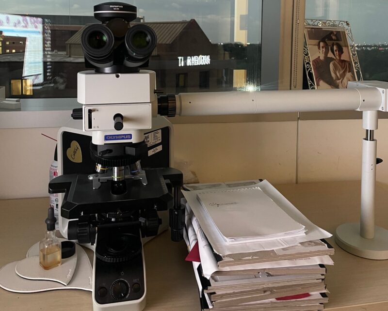
Sanam Loghavi shared on X/Twitter:
“Another day in the life of a hematopathologist….. To say I love this job is an understatement. Never a dull moment hemepathMDA…. Sharing some of the more photogenic cases we saw on service in this hemepath… in the order they appeared on my microscope. Let’s learn.
Relapsed AML (monoblastic), NPM1 mutated. A perfect example of why we still need the microscope for AML surveillance. Haven’t changed my stance since EHA2024 the flow sample was hemodiluted, with~5% monocytes showing slightly increased CD15 and partial CD56 (both non-specific). without microscope would be a very difficult ‘call’ by flow only but the BM is packed with blasts.

2. Acute promyelocytic leukemia with PML::RARA Immunophenotype: CD34 -, CD117 +, HLA-DR -, MPO uniformly + (remember this for your Boards) Note the ‘faggot cells’ with multiple Auer rods (arrow) on the aspirate. The promyelocytes on the HE (core bx) have abundant pale cytoplasm.

3. Hairy cell leukemia (HCL) flow: CD19/CD20/CD22 bright, CD11c +, CD103+, CD25+ PB: monocytopenia is typical Note the ‘hairy cells’ with circumferential cytoplasmic projections. Nucleoli are a ‘no no’ for HCL. On the core biopsy: note the typical ‘fried egg’ appearance with centrally located oval nuclei surrounded by pale cytoplasm. CD20 shows extensive BM involvement by HCL IHC if pos for BRAF V600E.

4. Acute myeloid leukemia Hx: ovarian cancer, s/p-platinum-based chemo. Progressive neutropenia.
NGS pending, but p53 IHC looked wild-type… so hopefully not a ‘therapy-related’ AML
Remember genetics trump history of therapy. My guess: this will be IDHm because the most significant cytopenia in this patient was neutropenia (will report back)..

5. Another NPM1 mutated AML… this time with perfect cup-like nuclei and NPM1 IHC with nuclear and cytoplasmic staining (cytoplasmic: abnormal and with the exception of rare NPM1 translocations, nearly always associated with NPM1 mutation) … my all-time favorite stain (fine… p53 is another one).

6. yet another NPM1 mut AML (not the 2nd most common mutation in AML 4 nothing)
Here blasts are not typical cup-like.. but phenotype suggestive of NPM1 mut: CD34-, CD117+, CD13 decreased, HLA-DR pos (HLA-DR may also be neg in NPM1).
NPM1 IHC is a slam dunk (nuclear +cytoplasmic staining)…
Look, I agree.. NPM1 may not be the prettiest stain out there, but what it lacks in beauty, it makes up in precision and skills…consider it the Steve
Buscemi of immunostains.

7. Patient with pancytopenia and relative monocytosis. I think it may be an evolving CMML… but for now does not meet the absolute monocyte count (it’s <500).
Why do I think CMML? BM monocytosis and ratio of classical monocytes to non-classical monocytes: 99.8% for now, best classified as MDS, NGS pending. I suspect a CMML-like mutation profile (TET2, SRSF2, etc)
I shall wait… and will report back to you guys, too… stay tuned.
Also… did you notice the plasma cells??? Incidental MGUS is hiding in here, too. PCs are aberrant by flow (CD19-, CD56+, lambda restricted).

That’s the end, friends. thanks for tuning in.. and I hope you learned something new.”

Source: Sanam Loghavi/X
Sanam Loghavi is an Associate Professor of Pathology and Laboratory Medicine at the Department of Hematopathology at MD Anderson Cancer Center. She is the Medical Director of the ECOG/ACRIN Leukemia Bank at the Central Biorepository and Pathology Facility at MD Anderson.
She has authored around 200 peer-reviewed publications focused on hematologic neoplasms and is a co-investigator on several national and internationally-run clinical trials for hematologic malignancies. She serves on various committees including the HPATH Committee of the College of American Pathologists and the Education and Outreach Committees for International Clinical Cytometry Society.


