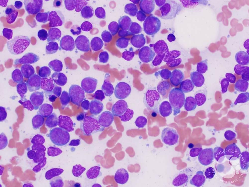
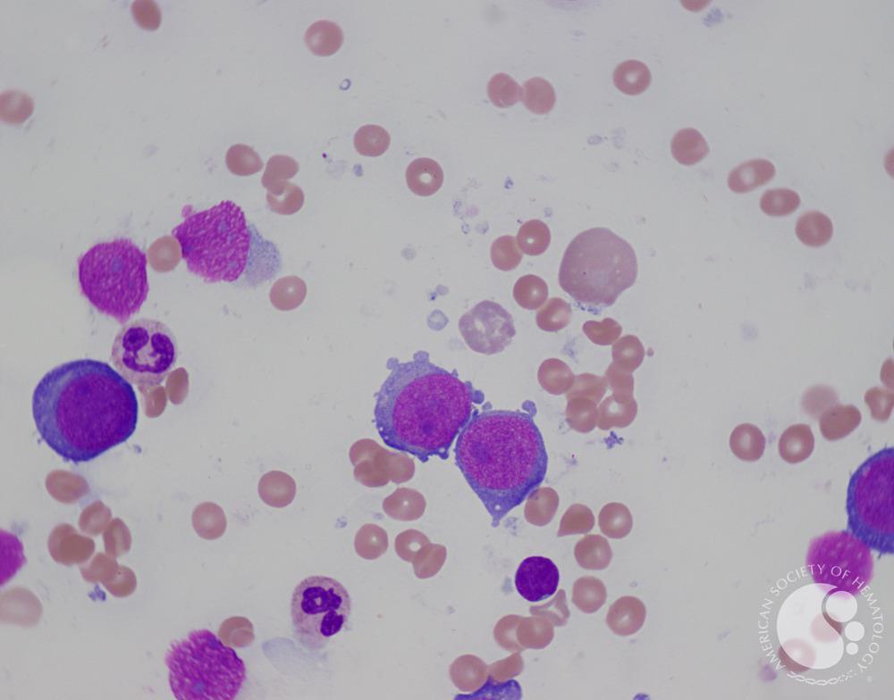
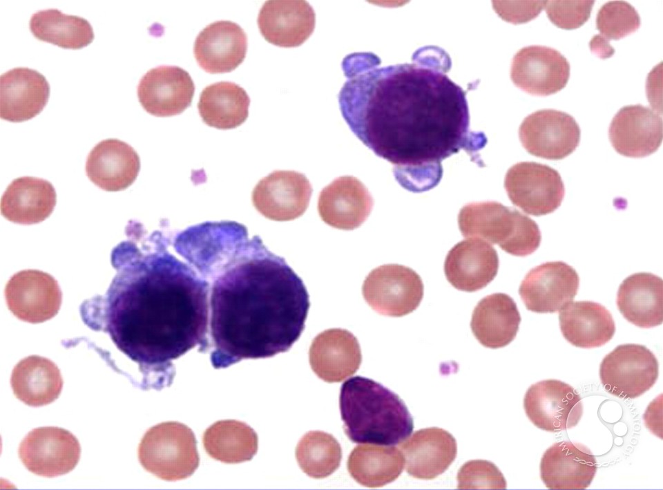
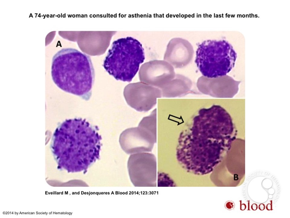
Nishant Rajendra Tiwari, Hemato-oncology fellow at OU Health Stephenson Cancer Center, shared a thread on X:
Educational thread on common microscopy and flow cytometry patterns for acute leukemias. Not medical advice. Always open for feedback and corrections. Blood cancer awareness month is September. Images are from ASH image bank!
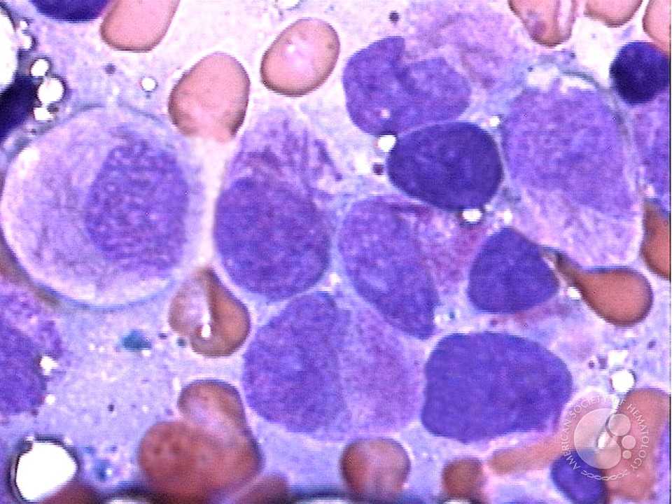
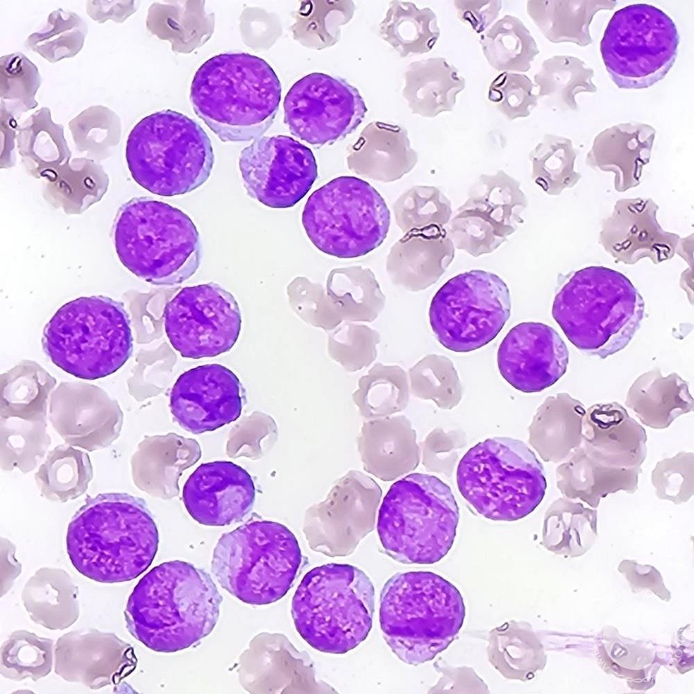
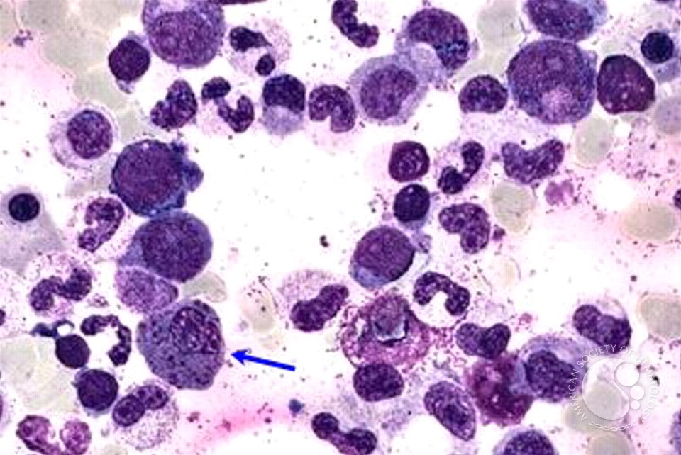
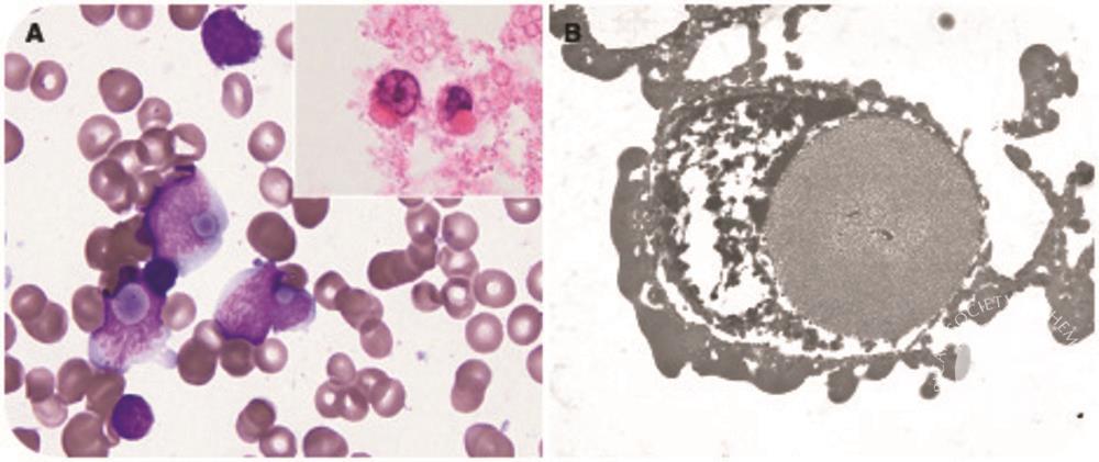
Acute promyelocytic leukemia (APL) hyper-granular variant –
Auer rods, Heavy granulation CD13 heterogenous, CD34-ve, CD33+, CD117+, MPO+ CD15-ve, CD11b-ve, CD11c-ve, CD15dim. Image from ASH image bank.
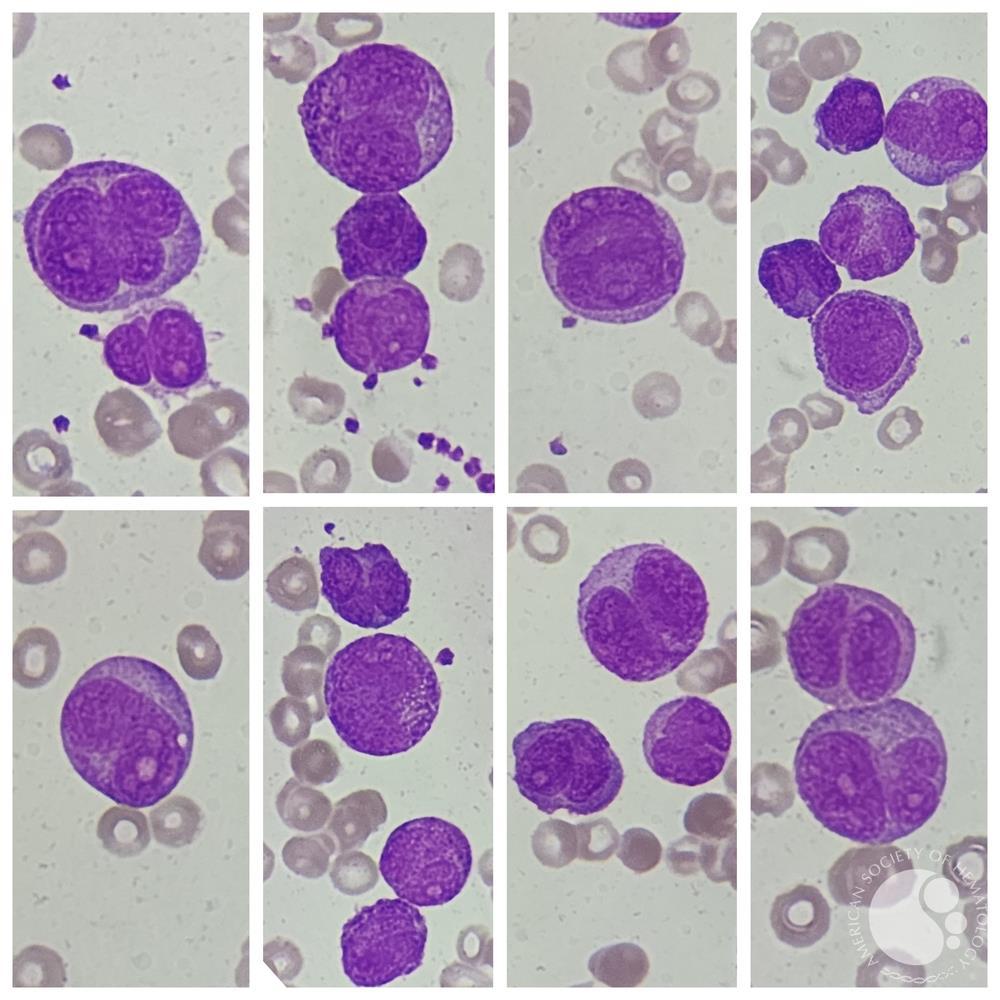
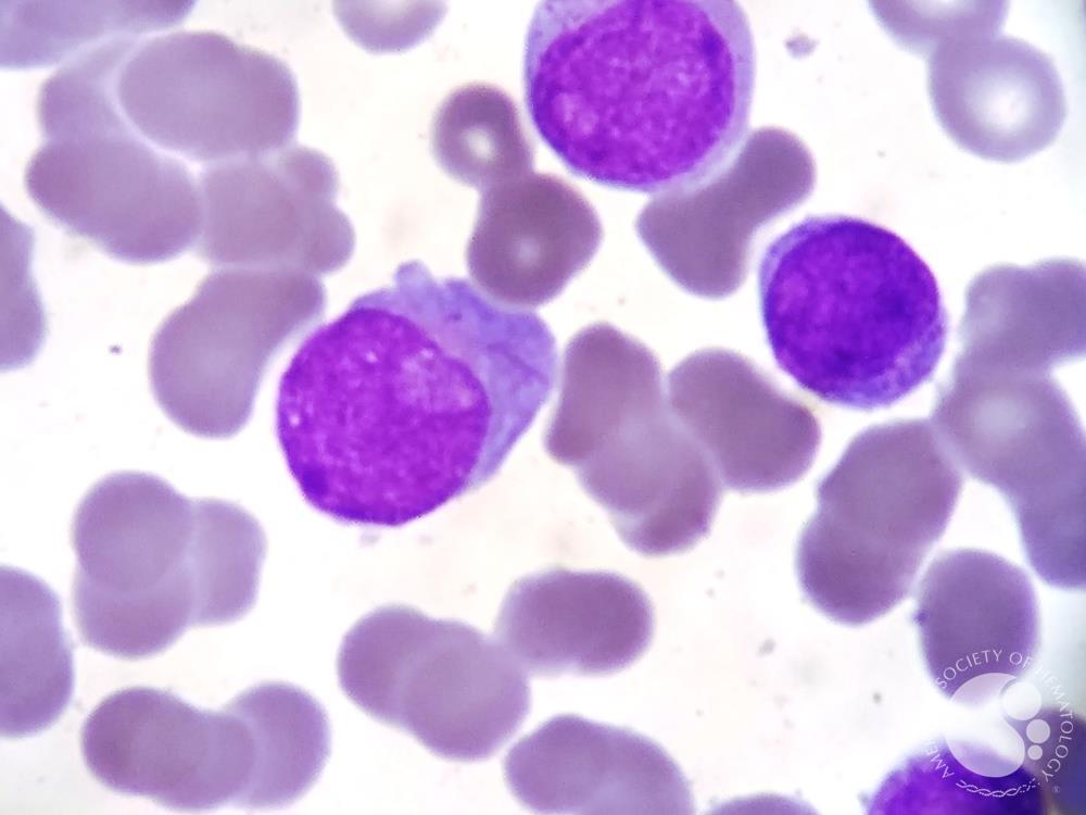
APL micro-granular variant –
Bilobed nucleus, paucity of/absent granules CD13+, CD34+ (often), CD33+, CD117+, CD2+, CD64+ CD15-ve, CD11b-ve, CD11c-ve, CD65-ve. Image from ASH image bank.
NPM1 mutated Acute myeloid leukemia (AML) –
Cup like blasts CD13+, CD33+, CD34-ve, CD`117+, HLD-DR-ve, CD110+, CD123+, CD15+
Images are from ASH image bank
AML with inv(16) or t(16::16) –
Abnormal eosinophils, eosinophilia CD13+, CD33+, CD34+, CD117+, HLA-DR+, CD11b+, CD11c+, CD15+, CD45+, CD2+, CD4+
Images are from ASH image bank
AML with t (8::21) –
Perinuclear Hofs, thin auer rods CD13+, CD33+ (weak), CD34+, MPO+, CD11b+, CD15+, HLA-DR (-/+), CD65+ CD56+ (Association with KIT mutation, worse prognosis)
Images are from ASH image bank
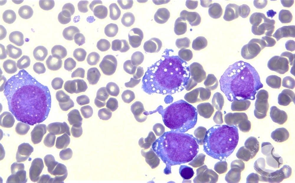
AML with t(9::11) –
Monocytic/moboblastic-cytoplasmic vacuolization CD13+, CD33+, CD14+, CD34-/+, HLA-DR+, CD38+, CD68+m CD64+, MAb 7.1+
Images are from ASH image bank
AML with t(6::9) –
Increased basophils CD13+, CD33+, CD117+, HLA-DR+ (In blasts-negative on basophils) CD34-ve at presentation CD34+ at relapse
Images are from ASH image bank
Acute erythroid leukemia –
Cytoplasmic blebs, basophilic cytoplasm CD13-ve, CD3-ve, CD117+, CD71+, Glycophorin+, hemoglobin+.
Images are from ASH image bank
Acute megakaryoblastic leukemia –
Cytoplasmic pseudopod formation, granular basophilic are in cytoplasm CD41+, CD61+, CD36+, CD42b+
Images are from ASH image bank
Acute basophilic leukemia –
Very rare, deep basophilic nuclei CD13+, CD33+, CD123+, CD11b+, CD117-ve CD34+/-ve, CD9+.
Images are from ASH image bank
B-cell acute lymphoblastic leukemia (ALL) with t(9::22) –R
ussell like bodies CD34+, CD19+, CD10+, CD20-/+, CD25+ CD15-ve, CD65-ve Often CD66c+, CD13+, CD33+
Images are from ASH image bank
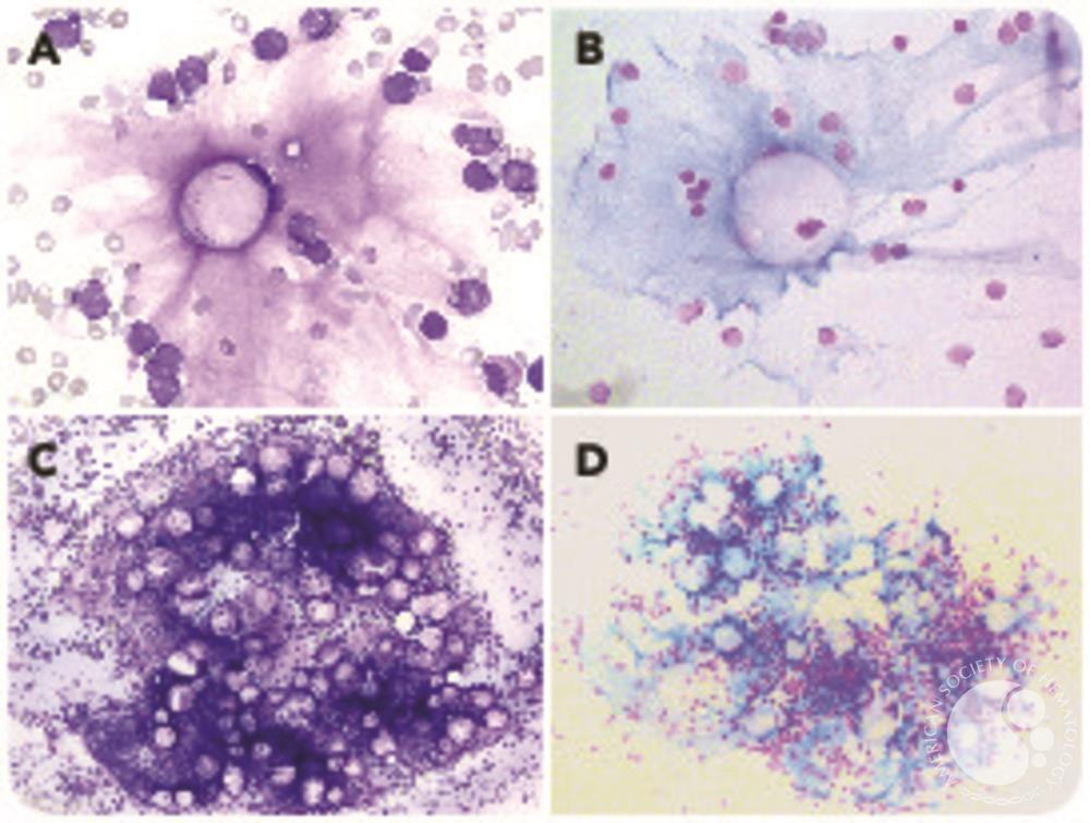
B-cell ALL with t(12::21) –
Gelatinous deposition in bone marrow CD34+/-ve, CD19+, CD10+, CD2-ve/+, CD13+, CD33+ CD66c-ve. CD15-ve, CD65-ve, MAb7.1 -ve, cytIgM-ve
Images are from ASH image bank
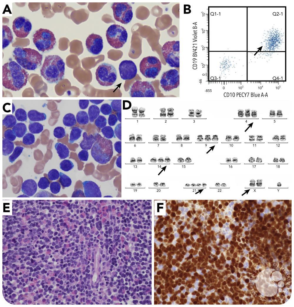
B-cell ALL with hyperdiploidy –
Hyper-eosinophilia CD34+, CD19+, CD10+, TdT+, CD123+, CD20-ve/+ CD13-ve, CD33-ve, CD15-ve. CD65-ve, MAb7.1 -ve, cytIgM -ve
Images are from ASH image bank
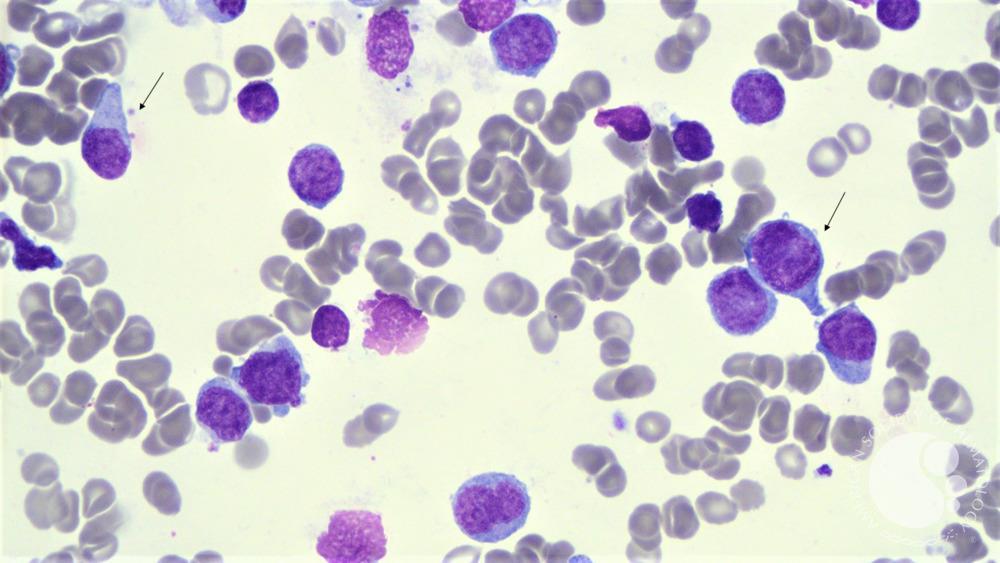
T-cell ALL –
Hand mirror confirmation of cells (Arrow) CD34+, CD7+, CD3+, CD33+, CD38+, cytTdT+
Images are from ASH image bank
B-cell ALL with t(4::11) –
no image
CD34+, CD19+, CD10-ve, CD20-ve, MAb7.1 +, cytIgM -ve Often CD15+, CD65+, CD13+, CD33+
B-cell ALL with t(1::19)-
no image
CD34-ve, CD19-ve, CD10-ve, CD20+, cytIgM+, CD13-ve, CD33-ve, CD15-ve, CD65-ve
B-cell ALL with IGH-
CLRF fusion-no image CLRF over expression
T-cell ALL with FLT3 activating mutation–
no image
CD117+, CD135+
There are many nuances to these, also many variants, which are hard to explain on a short tweet. I tried best to keep it lucid and clear. Happy to hear any feedback or corrections.
References –
-Wintrobe’s Clinical Hematology
-Clinical hematology atlas –pathologyoutlines.com
-ASH image bank
Source: Nishant Rajendra Tiwari/X
Nishant Rajendra Tiwari is a Fellow at OU Health Stephenson Cancer Center. After starting his residency at Loyola Medicine-MacNeal Hospital, he pursued a specialization in Hematology and Oncology. During this period, he conducted research on thrombotic thrombocytopenic purpura (TTP) under the mentorship of Dr. Spero Cataland.


