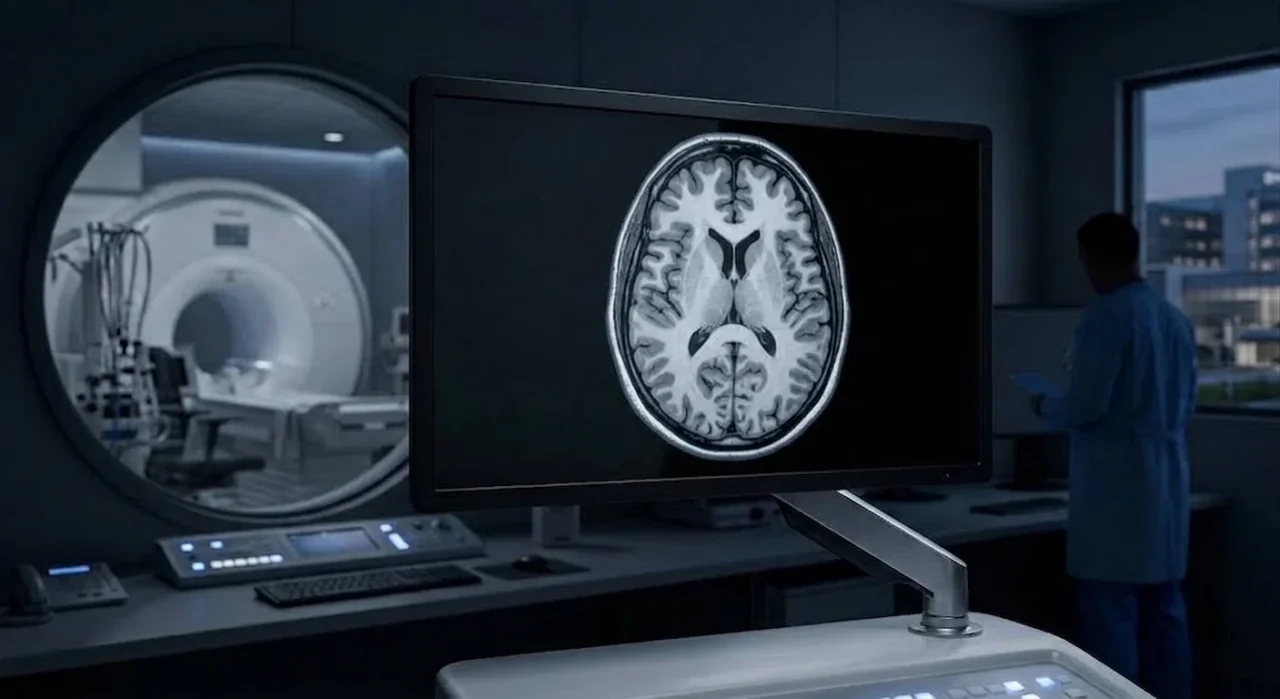
Researchers at The Royal Marsden NHS Foundation Trust have shown that routine whole-body MRI scans could reveal far more about a patient’s health than previously thought. Using a new artificial intelligence (AI) tool to analyse changes in muscle, body fat and organ volume, researchers found the scans could help predict how well patients with multiple myeloma respond to treatment.
The study, published in the Blood Advances journal, is the first to show that AI can automatically analyse healthy tissue on whole-body MRI scans performed as part of routine myeloma care. The technique provides new imaging biomarkers that could help identify patients at greater risk of disease progression, without requiring any additional scans or hospital appointments.
Multiple myeloma is an incurable cancer of the plasma cells, with around 6,000 people diagnosed every year in the UK. Patients with multiple myeloma often undergo prolonged treatment involving combinations of drugs over several years. While advances in treatment have significantly improved survival, they can also affect muscle, body fat and overall physical health. Until now, clinicians have had limited ways of measuring these changes objectively.
Researchers at The Royal Marsden developed an AI tool capable of automatically measuring skeletal muscle, body fat and organ volumes from routine whole-body MRI scans. They applied the technology to scans from patients taking part in a prospective study, who underwent imaging; before treatment, after induction chemotherapy, and following a stem cell transplant.
The researchers found that treatment was associated with measurable changes in body composition. Abdominal skeletal muscle decreased following induction chemotherapy, while both subcutaneous and visceral fat increased during treatment before partially recovering after a stem cell transplant.
Subcutaneous fat is the soft fat stored just beneath the skin, whereas visceral fat is the deeper fat stored around internal organs.
Importantly, these changes were also linked to patient outcomes. Patients with higher levels of abdominal skeletal muscle and subcutaneous fat at diagnosis experienced longer progression-free survival. Increases in visceral fat during treatment were associated with a greater risk of disease progression. When combined with routine clinical information, the AI-derived measurements accurately predicted patient outcomes, demonstrating their potential value for improving risk assessment.
The findings suggest body composition analysis could become an important addition to routine MRI assessments, helping clinicians identify patients who may benefit from earlier supportive interventions such as nutritional support, exercise programmes or physiotherapy, as part of a more holistic and personalised approach to cancer care.
Unlike conventional measures such as body mass index (BMI), AI-derived body composition measurements provide detailed information about the distribution of muscle and fat throughout the body, offering a more accurate picture of a patient’s overall physical health and resilience during treatment.
The study also highlights how AI can unlock additional value from scans that patients already receive as part of standard care. While manually analysing whole-body MRI scans can take several hours per patient, the automated AI tool performs the same analysis in just minutes, making it practical for larger-scale clinical use.
Professor Christina Messiou, Consultant Radiologist at The Royal Marsden, Professor in Imaging for Personalised Oncology at The Institute of Cancer Research, London and Chief Investigator of the study, said:
“This research shows that we can learn far more from the MRI scans that patients are already having as part of their care. By using AI to understand changes in body composition over the course of treatment, we hope to build a more complete picture of a patient’s overall health, not just how their cancer is responding. In the future, this approach could help us identify patients who would benefit from a more holistic approach to cancer care, including additional support such as physiotherapy, nutritional advice or exercise programmes, and may even allow us to detect treatment side effects earlier. It could also help us compare the impact of different therapies on patients’ physical wellbeing, alongside their effectiveness against the cancer. While this technology is still at the research stage, our long-term vision is for every whole-body MRI scan to provide not only information about the disease itself, but also valuable insights into a patient’s overall physical health, helping us deliver more personalised care.”
Researchers say larger, multicentre studies are now needed to confirm these findings before the technology can be introduced into routine clinical practice. If validated, AI-powered body composition analysis could provide clinicians with a simple, scalable and non-invasive way to improve risk assessment while making better use of scans that patients already receive as part of their care.
This research was supported by, The Royal Marsden Cancer Charity, the National Institute for Health and Care Research (NIHR) Biomedical Research Centre at The Royal Marsden NHS Foundation Trust and The Institute of Cancer Research, London, and Cancer Research UK’s National Cancer Imaging Translational Accelerator (NCITA).
You can also read:
Shaalan Beg: When AI Enters the Clinic

Myeloma Paper of the Day, September 13th, Suggested by Robert Orlowski