





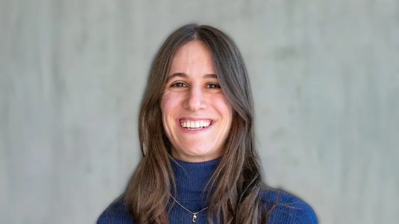
Lydia Schönpflug, PhD Researcher in Computational Pathology at University Hospital Basel and University of Basel, shared on LinkedIn:
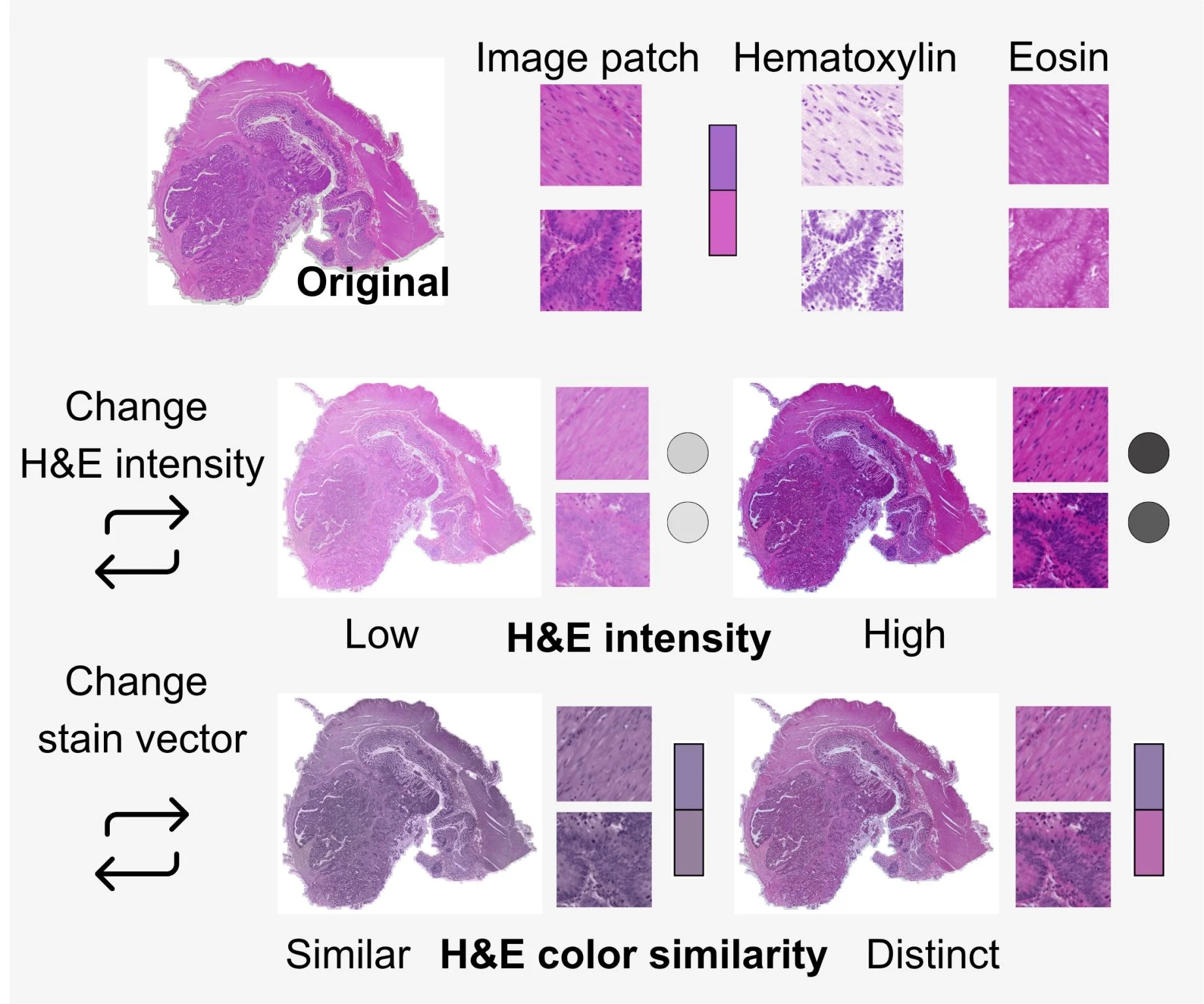
“Same tumor, same slide scanned in two different hospitals: One comes out pale pink. The other, deep purple.
That’s H&E staining variation, and most AI models in computational pathology have not been tested against it systematically.
We just published a protocol that adresses that gap.
We simulate changes in staining intensity and color built on quantifiable targets, derived from optical density stain vector decomposition for both source and target staining. As far as we know, it’s the first approach to define measurable staining targets rather than just picking a reference image or physically rescanning/restaining the slide.
As a test case, we ran it on 306 models detecting microsatellite instability in colorectal cancer on the SurGen dataset (n=738 WSIs). The finding that stuck with us: high accuracy does not necessarily equate high robustness to staining shifts (r~0.28), making an independent measurement of robustness essential.
Big thank you to my co-authors and collaborators Maxime Lafarge, Viktor H. Koelzer, Nikki van den Berg, Tjalling Bosse, Jurriaan Barkey Wolf, Sonali Andani, Nanda Horeweg.”
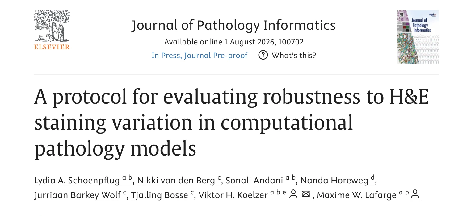
Title: A protocol for evaluating robustness to H&E staining variation in computational pathology models
Authors: Lydia A. Schoenpflug, Nikki van den Berg, Sonali Andani, Nanda Horeweg, Jurriaan Barkey Wolf, Tjalling Bosse, Viktor H. Koelzer, Maxime W. Lafarge
Read the Full Article.
Other articles about Computational Pathology on OncoDaily.