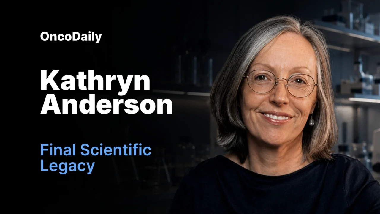
More than five years after the death of developmental biologist Kathryn Anderson, PhD, researchers at Memorial Sloan Kettering Cancer Center have completed and published the final study originating from her laboratory.
Published on July 1, 2026, in Developmental Cell, the study examines how WNT signaling guides highly flexible embryonic cells toward distinct and specialized identities. The findings provide new insight into early mammalian development while raising important questions about how similar molecular programs may contribute to cancer metastasis.
The paper, titled “AXIN1 and AXIN2 regulate the WNT-signaling landscape to promote distinct mesoderm programs,” represents both a major scientific contribution and the culmination of years of work by Dr. Anderson’s former colleagues and collaborators.
A Final Chapter in a Landmark Scientific Career
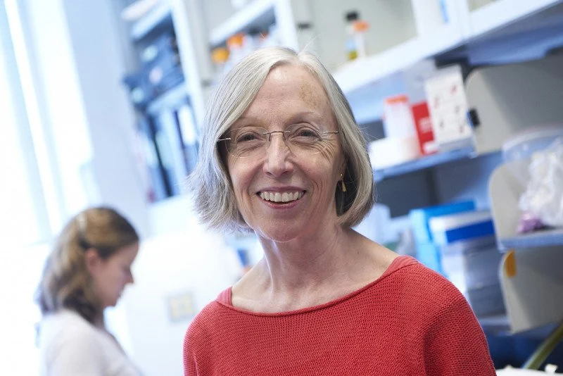
Dr. Anderson joined Memorial Sloan Kettering Cancer Center in 1996 and later served as Chair of the Developmental Biology Program. Her research focused on the fundamental processes that shape early mammalian development, including how highly plastic embryonic cells receive instructions that determine which tissues and organs they will ultimately form.
She died in November 2020 after a career marked by major discoveries in developmental biology.
The research leading to the new publication began approximately a decade ago in Dr. Anderson’s laboratory. Following her death, the project was continued by former laboratory members and colleagues who were determined to bring the work to completion.
Anna-Katerina Hadjantonakis, PhD, who succeeded Dr. Anderson as Chair of the Developmental Biology Program at MSK, oversaw the research during its later stages.
The Mutation That Opened a New Research Path
The project originated when Rocio Hernández-Martínez, PhD, then a postdoctoral researcher in Dr. Anderson’s laboratory, helped develop a mouse model lacking two closely related genes, Axin1 and Axin2.
Under normal conditions, these genes help regulate WNT signaling and prevent the pathway from becoming excessively active. When both genes were absent, WNT signaling remained continuously switched on.
The resulting embryos developed profound abnormalities. They were able to produce only a limited range of tissues and lacked cells that would normally contribute to the heart, head, and other structures located toward the front of the body.
The unusual developmental pattern provided researchers with an opportunity to investigate how WNT controls the earliest decisions made by embryonic cells.
Mapping the First Decisions of Cell Development
For the new study, researchers developed genetic tools that allowed them to deactivate the Axin genes specifically within the epiblast.
The epiblast is a thin layer of highly plastic cells that gives rise to nearly every tissue in the body. At this early stage, the cells retain the potential to develop into many different cell types.
The researchers combined these genetic tools with single-cell sequencing, allowing them to examine gene activity within individual cells and study how cells respond to signals from their surroundings.
This approach enabled the team to map, in considerable detail, how WNT influences the transition from cellular plasticity to specialized identity.
WNT Does Not Deliver a Single Instruction
The study found that WNT signaling acts through multiple stages rather than producing one uniform effect.
Initially, WNT pushes epiblast cells away from their highly flexible state and begins directing them toward the mesoderm, the embryonic tissue that later contributes to muscles, bones, organs, blood vessels, and connective tissues.
However, WNT alone does not determine the final identity of a cell.
Instead, the eventual outcome depends on how WNT interacts with additional molecular signals across the developing embryo.
“WNT has multiple roles,” Dr. Hadjantonakis explained. “It’s pushing cells from one state to the other initially, and then it’s integrating two distinct molecular signals across a spatial landscape to define the final outcome.”
A Molecular Map Defined by BMP and NODAL
The researchers identified two particularly important signals involved in this process: BMP and NODAL.
Both belong to the transforming growth factor-beta, or TGF-beta, family of signaling molecules. Despite belonging to the same family, they direct cells toward different developmental outcomes.
BMP and NODAL form opposing gradients across the embryo. BMP activity is associated with cell identities toward the back of the developing body, while NODAL signaling helps direct cells toward front-of-body structures.
Each embryonic cell effectively reads its position within these gradients and combines that information with WNT signaling to determine what type of cell it should become.
“BMP with WNT gives you one outcome; NODAL with WNT gives you another,” Dr. Hadjantonakis said.
The finding highlights an important principle in developmental biology: cellular identity is not determined by the presence of a single signal, but by the combination, strength, timing, and location of several signals acting together.
From Embryonic Development to Cancer Metastasis
The study may also have implications for understanding cancer metastasis.
To spread from a primary tumor to distant organs, cancer cells must separate from neighboring cells, become mobile, and travel through surrounding tissues. This behavior involves a biological program known as epithelial-to-mesenchymal transition, or EMT.
EMT is essential during embryonic development because it allows cells to move to the correct locations as the body forms. In cancer, however, similar cellular changes may support tumor invasion and metastasis.
TGF-beta signaling is already known to play an important role in driving EMT in cancer. The new study suggests that it may be insufficient to consider TGF-beta signaling as one uniform biological process.
Although BMP and NODAL are both members of the TGF-beta family, they operate through different molecular mechanisms and can direct cells toward opposing outcomes.
“Not all TGF-betas are the same,” Dr. Hadjantonakis said. “BMP with WNT gives you one outcome. NODAL with WNT gives you another.”
Understanding these distinctions may help researchers better define how WNT and TGF-beta-related pathways interact within tumors and contribute to the ability of cancer cells to spread.
Why the Identity of the Signal Matters
Metastasis remains one of the most serious challenges in cancer care and is responsible for the large majority of cancer-related deaths.
The study does not establish a new treatment for metastasis. However, it provides a more precise framework for examining the molecular signals that may enable cancer cells to change identity, detach from surrounding tissue, and migrate.
Rather than asking only whether TGF-beta signaling is active, future research may need to determine which member of the TGF-beta family is active, where the signal originates, and how it interacts with WNT and other pathways.
These distinctions could reveal new opportunities to interfere with the molecular programs that support tumor progression and metastatic spread.
Completing the Work Against the Odds
The study faced several significant obstacles before publication.
Dr. Anderson became ill and began supervising her laboratory remotely. Soon afterward, the COVID-19 pandemic disrupted research activities across the institution and made it more difficult to complete essential experiments.
Following her death, members of her laboratory moved into new research groups at MSK. Dr. Hernández-Martínez joined Dr. Hadjantonakis’ laboratory, where she continued working on the WNT project for another two years before accepting a position at the University of California, San Francisco.
With no dedicated funding and researchers balancing the study alongside their other responsibilities, completing the project became a collective effort.
Senior research scientist Sonja Nowotschin, PhD, and senior research assistant Ying-Yi Kuo, MS, conducted additional experiments needed to move the study toward publication. Researchers from the laboratory of Bertie Göttgens, DPhil, at the Cambridge Stem Cell Institute in the United Kingdom contributed essential expertise in genomics.
“It was everyone’s side project, but we were determined to see it through,” Dr. Hadjantonakis said.
A Scientific Commitment and a Personal Tribute
For the researchers involved, completing the paper was about more than publishing scientific results.
Dr. Hernández-Martínez described Dr. Anderson as an engaged and curious mentor who regularly visited the laboratory to discuss experiments and ask researchers about their projects.
“Completing this project was the best way to honor her,” she said.
Dr. Hadjantonakis similarly described the work as both a scientific obligation and a personal commitment to a friend and colleague.
The study preserves Dr. Anderson’s final contribution to a central question in developmental biology: how cells interpret several competing signals and transform that information into a clear developmental decision.
New Questions at the Intersection of Development and Cancer
The publication also creates new directions for research.
Scientists must now investigate how WNT integrates with BMP and NODAL signals at the molecular level and how these interactions change across different tissues and biological settings.
In both embryos and tumors, cells exist within complex three-dimensional environments where multiple signals are active simultaneously. Each cell must determine which signals to respond to, which to ignore, and how those signals should influence its behavior.
In embryonic development, this process is highly organized and allows tissues and organs to form in the correct locations. In cancer, similar signaling pathways may become disrupted, enabling cells to acquire abnormal identities and invasive properties.
By revealing how WNT, BMP, and NODAL work together to guide embryonic cell identity, the study brings researchers closer to understanding how these same pathways may be altered during cancer progression.
The research was supported by grants from the National Institutes of Health, the Wellcome Trust, the Pew Latin American Fellows Program, and the National Cancer Institute Core Grant supporting Memorial Sloan Kettering Cancer Center.
Additional authors included Luke T.G. Harland, Bart Theeuwes, and Elizabeth Lacy.
Study: Hernández-Martínez R, et al. “AXIN1 and AXIN2 regulate the WNT-signaling landscape to promote distinct mesoderm programs.” Developmental Cell. Published July 1, 2026. DOI: 10.1016/j.devcel.2026.06.004.
Written by Nare Hovhannisyan, MD
Stay informed with more articles like this on OncoDaily.