
C. Ola Landgren, Leader, Translational and Clinical Oncology Program/Experimental Therapeutics at Sylvester Comprehensive Cancer Center, shared on LinkedIn:
“As treatments for multiple myeloma become more effective, our ability to see the disease must advance alongside them.
Today, our new phase II study, published in Radiological Society of North America (RSNA) journal Radiology, marks an important step toward developing targeted imaging in parallel with targeted therapies in myeloma.
For decades, skeletal surveys were used to detect myeloma bone disease. Yet substantial local bone destruction, often approximately 50% or more, can occur before a lesion becomes visible on X-ray.
Whole-body MRI allows assessment of bone and bone marrow, including functional information through diffusion-weighted imaging. Access and specialized interpretation remain limited in many settings.
FDG-PET/CT is another important advance. Its CT component identifies bone destruction, while PET measures glucose uptake. However, some myeloma lesions show little increased FDG uptake. Our earlier work described negative FDG imaging in approximately 30% of patients with established myeloma bone disease. Conversely, increased FDG uptake can occur with inflammation, degenerative joint disease, infection, and trauma.
These limitations motivated us to develop CD38-targeted immunoPET: a radiolabeled antibody designed to recognize CD38 on myeloma cells.
We developed the preclinical work in collaboration with Dr. Jason Lewis at Memorial Sloan Kettering Cancer Center and Dr. Eric Smith, now at Dana-Farber Cancer Institute. This laboratory work led to our first-in-human phase I study, published in 2020, establishing safety, dosing and imaging timing. Next, my longtime collaborator Dr. Gary Ulaner, now at Hoag Cancer Center, and I developed this phase II study to compare CD38-targeted immunoPET against standard-of-care FDG PET/CT.
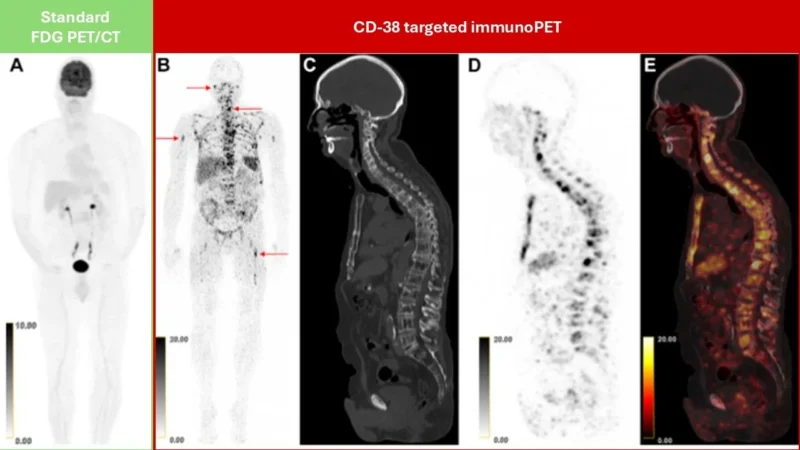
In our prospective study of 60 patients, immunoPET identified more disease sites than FDG-PET/CT in 28% of participants. In eight patients, disease was visible only on immunoPET, and targeted biopsies confirmed active myeloma in all eight. One striking example revealed more than 100 bone foci despite a negative FDG-PET scan (see below).
The approaches showed complementary strengths. ImmunoPET also requires careful interpretation, but these findings demonstrate that targeted imaging can reveal myeloma that conventional imaging misses.
This work is part of our broader initiative to develop curative therapies for myeloma. Developing targeted imaging alongside targeted treatments may help us understand where disease persists and how best to address it.
Next, we aim to develop improved tracers, investigate residual-disease assessment, and test whether imaging-guided decisions improve outcomes. Grateful to our patients and multidisciplinary teams. Together, we are moving the needle!”
You can also read: Early Relapsed Myeloma: No Efficacy Hierarchy Established Between CAR T Cells and Bispecific Antibodies