“Welcome to Day 7 of our Spir Takeover series featuring Jurgen Runge!
Today, we stay in the realm of paediatric interventional oncology and explore radioembolization and MWA of the liver. Whilst both techniques have a clear role in the treatment of adult patients, their role needs to explored and defined more clearly in the paediatric setting. Nevertheless, in Utrecht we have seen a slow increase in numbers, from 1 case in 2020, to 6 in 2024.
Radioembolisation
An adolescent with residual liver metastases post-chemotherapy was considered for radioembolization and microwave ablation (MWA). A test-treatment using DSA and conebeam-CT (CBCT) helped assess hepatic arterial anatomy and plan injection positions.
DSA revealed a hypertrophied right gastric artery (Fig. 1), which could lead to extrahepatic deposition if a central injection position was used. CBCT confirmed bilobar liver metastases (Fig. 2). MAA was injected to assess pulmonary shunt and extrahepatic deposits, which were confirmed in the stomach and spleen on MAA-SPECT/CT (Fig. 3).
Treatment involved MWA for left-sided lesions and a specific S5/8 lesion (Fig. 4), while Y90 was selectively injected into the S5/8 branch and the main right hepatic artery, with excellent targeting confirmed by Yttrium PET/CT (Fig. 5-8).
Liver MWA – HepACAGA
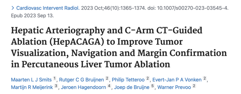
Liver MWA is typically performed using the HepACAGA technique, which involves hepatic arteriography and C-arm CT-guided ablation. In this approach, the patient remains in the angiosuite and CBCT hepatic arteriography is done in apnoea, eliminating the need for patient transfer.
The trajectory for MWA antenna insertion is planned and monitored fluoroscopically using needle guide software. This technique is demonstrated in a case of ablation in a young teenager (Fig. 1-2). The advantage of staying in the angiosuite is that, in case of hemorrhage, immediate DSA and embolization can be performed without moving the patient.
Liver MWA – US
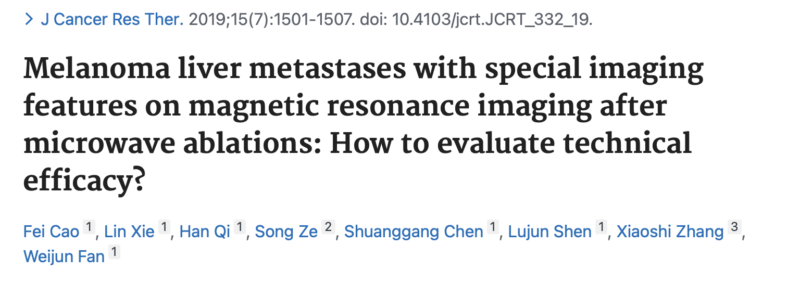
While HepACAGA is the primary technique used for liver MWA in Utrecht, ultrasound (US) is sometimes employed for quick needle placement, especially in small lesions. A case of solitary melanoma metastasis in a teenager showed typical imaging features, including native T1W hyperintensity (Fig. 1) and diffusion restriction (Fig. 2), which were visible on US (Fig. 3).
Post-ablation, the lesion did not show enhancement or diffusion restriction, which was interpreted as complete ablation. Persistent T1W hyperintensity after ablation, commonly seen in melanoma metastasis, does not indicate incomplete ablation, as noted in previous studies by Cao et al.
Cases performed by: Rutger Bruijnen and Maarten Smits
Please mark your agendas for the paediatric IO clinical focus (CF212) session at ECIO2025 in Rotterdam, the Netherlands on Monday April 14th 2025!”
References:
Authors: Maarten Smits et al.

Authors: Fei Cao et al.

Proceed to the video attached to the post.
Dr. Jurgen Runge, MD, PhD, is a Fellow in Pediatric and Interventional Radiology at UMC Utrecht’s Wilhelmina Children’s Hospital. Prior to fellowship, he was an Interventional Radiologist at the Antoni van Leeuwenhoek Hospital.
Dr. Runge completed his radiology residency at Amsterdam UMC and OLVG, the primary city hospital of Greater Amsterdam. His current research interests focus on pediatric interventional radiology and pulmonary ablation.
