For decades, cancer researchers have hit a fundamental roadblock before a drug ever reaches a patient: the limitations of a flat petri dish. Traditional cancer research grows cells from a patient’s tumor in two dimensions, flattened onto plastic plates. The approach has produced important breakthroughs, but it does not recreate reality. Human tumors don’t grow as flat, isolated sheets. They are complex three-dimensional masses embedded within a living tapestry of blood vessels, immune cells and structural proteins.
This architectural discrepancy is especially problematic in head and neck squamous cell carcinoma (HNSCC). Head and neck cancers are notoriously heterogeneous, meaning cells within a single tumor can differ. These tumors are also strongly shaped by the microenvironment, the dense, fibrotic and highly specific neighborhood of tissue in the tongue, throat or jaw. When cells are flattened onto plastic, they behave differently and lose their native characteristics, and drugs that appear effective against them frequently fail in human clinical trials (Tuveson and Clevers, 2019).
Structural engineering is biotechnology’s most recent recruit for filling this gap. Decellularization and 3D bioprinting let researchers build
The Architectural Blueprint: Decellularized Matrices
Before you can print a realistic tumor, you need the right canvas.
This is where decellularized extracellular matrix (dECM) technology becomes essential. The extracellular matrix (ECM) is the non-cellular network of structural proteins such as collagen, laminin and fibronectin that gives tissues their mechanical shape and physical stiffness.
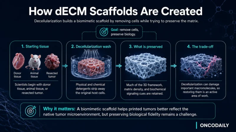
To create a biomimetic scaffold, scientists take donor tissue, animal tissue or resected tumor and process it with physical and chemical detergents. This washing step strips away the original host cells while preserving much of the underlying 3D framework, matrix density and biochemical signaling cues. The trade-off is that decellularization is disruptive: it can damage the very macromolecules that make the scaffold biologically meaningful, and restoring them is an active area of work (Siahmansouri et al., 2026).
Recreating this native matrix still matters for HNSCC modeling. Head and neck tumors rely on the stiffness and extracellular architecture of surrounding oral tissues to drive local invasion and develop drug resistance. By repopulating a ghost-like dECM scaffold with patient-derived HNSCC cells, the cancer cells inhabit an environment that reflects the stiffness and signaling of human tissue far more closely than plastic ever could, encouraging them to cluster, migrate and respond to chemotherapy more as they would inside a patient.
Printing the Tumor Microenvironment
Decellularized scaffolds provide the backdrop, but 3D bioprinting lets scientists recreate tumor architecture with microscopic precision. Instead of plastic or metal, a 3D bioprinter uses “bio-inks”: hydrogels loaded with living cells.
The bioprinter’s robotic nozzles deposit layers of bio-ink according to a computer-generated blueprint, sometimes informed by tissue imaging, though most current models use defined shapes such as droplets and cylinders to optimize cell survival.
Researchers are not only printing cancer cells, but other components of the tumor microenvironment as well. Stromal fibroblasts have been co-printed with tumor cells in dECM-based bio-inks, producing constructs with a cancer cell core and a fibroblast periphery that model the protective matrix barriers defending head and neck tumors against chemotherapy (Kort-Mascort et al., 2023). Endothelial cells, which form the primitive vessels a tumor uses to recruit its blood supply, and immune cells remain next targets rather than routine inclusions (Azhakesan et al., 2025).
Printing distinct cell types into precise spatial arrangements is what will eventually allow an accurate living model of one patient’s disease.
Shifting from Generalized to Personalized Medicine
The end game of this work is to take the guesswork out of cancer treatment. At present, a patient with advanced head and neck cancer is treated with established, aggressive protocols: a heavy combination of surgery, radiation and chemotherapy.
Bioprinting offers a route to rapid, personalized drug screening. A small biopsy taken at diagnosis can be expanded in culture and bioprinted into many near-identical miniature 3D tumors. Rather than trying untested regimens on the patient, researchers can evaluate different chemotherapy and targeted drug combinations on these models in parallel. Bioprinted HNSCC constructs seeded with patient-derived cells have been used to test radiochemotherapy, producing responses closer to those seen in patients than spheroid models did (Azhakesan et al., 2025).
This remains preclinical. Expanding enough cells from a small biopsy is a bottleneck, and immunotherapy screening will require models that contain immune cells. But the approach could eventually let clinicians choose an optimized regimen from day one.
The Horizon of Biotech Oncology
Rebuilding head and neck tumors in 3D represents a genuine shift in oncology, moving cancer research from a flat abstraction toward a tangible, spatial reality. By modeling the structural barriers and cellular relationships of HNSCC, biotechnology is giving scientists a clearer view of the enemy.
Perfusion systems that push fluid through printed constructs to simulate blood flow are already emerging, and standardization across labs is the next hurdle. The petri dish served its purpose for the last century, but the future of cancer therapy is three-dimensional.
Written by: Tatev Barseghyan
Read more biotech insights on OncoDaily Biotech.