
Thomas Hope, Professor in Residence at University of California, shared on LinkedIn:
“Thank you Derek Johnson for leading the Society of Nuclear Medicine and Molecular Imaging (SNMMI) appropriate use criteria for FET PET, just published in The Journal of Nuclear Medicine – JNM.
This document will guide appropriate use and help support reimbursement as access expands. It was a pleasure to work with this group and to learn from our European colleagues as well as neurosurgeons, radiation oncologists and neuro-oncologists.
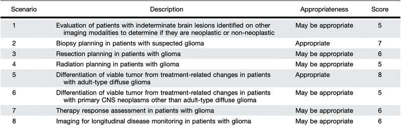
Two indications were rated appropriate:
- Biopsy planning in patients with suspected glioma.
- Differentiating viable tumor from treatment-related changes in adult-type diffuse glioma.
With FET PET approved, it will be interesting to see how it is adopted in clinical practice and where the evidence takes us next.”
To which, Derek Johnson, Chair of Division of Nuclear Medicine at Mayo Clinic, added:
“Thanks for the shout-out, Tom!
Looking into my crystal ball, I think some of the most important future uses of FET fell into the intermediate ‘May Be Appropriate‘ category in the present AUC.
There is a lot of work still to be done in this field!”
Title: Appropriate Use Criteria for 18F-Floretyrosine (FET) PET Imaging in Patients with Primary Brain Tumors
Authors: Derek R. Johnson, Nathalie L. Albert, William G. Breen, Shawn L. Hervey-Jumper, Raymond Y. Huang, Jana Ivanidze, Michelle M. Kim, Jonathan E. McConathy, Nancy Ann Oberheim Bush, Ian F. Parney, Matthias Preusser, Roger Stupp, Erik P. Sulman, Michael C. Veronesi, Javier E. Villanueva-Meyer, Thomas A. Hope.
More About
Pixclara (18F-floretyrosine) is an FDA-approved PET Radiotracer used to image brain tumors. It is a radioactive-labeled amino acid analog (fluorine-18) that enters glioma cells more rapidly than normal brain tissue by binding to LAT1 and LAT2 (L-type amino acid transporters).
Older nuclear medicine tools such as 18F-FDG PET never achieved broad clinical adoption in neuro-oncology due to high background glucose uptake in normal brain tissue. FET offers superior tumor-to-background contrast compared to FDG and does not require an on-site cyclotron like 11C-methionine.
FET PET is exceptionally well tolerated. Clinical trials and literature show a very low incidence of adverse events. The primary reported side effects are minor (headache at ≥ 0.5%, with nausea, injection site reactions, fatigue, and malaise occurring at <0.5%).Therefore, the primary clinical risk is not direct drug toxicity, but rather the potential for misinterpretation to negatively influence patient management decisions.
To standardize practice, the document aligns with international guidelines, recommending an adult dose of 5 to 7 mCi (185 to 259 MBq) with a 20-minute static PET acquisition starting 20 to 30 minutes after injection.
While dynamic PET imaging can provide additional kinetic data to help separate tumor progression from treatment effects, it remains largely confined to research settings due to scanner scheduling constraints. The scans can be performed on either PET/MRI or PET/CT systems, provided PET/CT images are fused with a recent anatomical MRI and interpreted by specialists experienced in multimodality glioma imaging.
The document focuses exclusively on primary brain tumors, specifically adult-type diffuse gliomas defined by the World Health Organization (WHO) classification:
- Glioblastoma, IDH-wildtype
- Astrocytoma, IDH-mutant
- Oligodendroglioma, IDH-mutant (and 1p/19q-codeleted)
The multidisciplinary panel of experts developed these criteria to establish formal, evidence-based recommendations on when and how FET PET should be ordered in clinical practice. The primary goal is to optimize diagnostic decision-making, particularly when evaluating treatment response versus progression, while preventing misuses that could negatively impact patient management.
You can also read: FDA Approves Floretyrosine F 18 for PET Imaging in Glioma