Small cell lung cancer remains one of the most difficult malignancies in which to generate durable benefit from immune checkpoint inhibition.
The paradox is familiar. SCLC is biologically aggressive and genomically complex, yet its responsiveness to immunotherapy remains substantially more limited than might be expected from tumor mutational burden alone. Platinum-based chemotherapy combined with an immune checkpoint inhibitor is established first-line therapy for extensive-stage disease, but durable responses remain uncommon. The authors estimate that approximately 80%–90% of patients do not achieve durable benefit from immunotherapy.
A new study in Nature Communications proposes a mechanistic explanation for part of this immune resistance, and identifies a potentially druggable pathway connecting tumor-cell nuclear export, macrophage polarization, antigen presentation and response to PD-1 blockade.
The central player is Exportin 1 (XPO1).
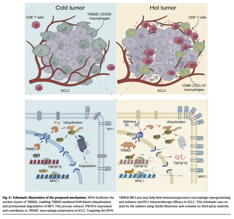
Using SCLC cell models, single-cell transcriptomics, murine tumors and patient-derived samples, investigators describe an XPO1–TRIM21–IRF3–TNFSF15–TREM2 pathway through which tumor cells appear to reshape the surrounding immune microenvironment.
Pharmacologic or genetic inhibition of XPO1 reduced immunosuppressive TREM2-positive macrophages, restored MHC class I expression and CD8-positive T-cell infiltration, and enhanced the antitumor activity of PD-1 blockade in preclinical models.
The findings are translationally intriguing. But the distinction is essential, this is a mechanistic and preclinical study, not evidence that XPO1 inhibition improves immunotherapy outcomes in patients with SCLC. That question now requires clinical testing.
Why SCLC Remains Difficult to Immunologically Engage
One important feature of SCLC immune resistance is impaired antigen presentation.
When tumor cells express insufficient MHC class I, cytotoxic T cells have fewer opportunities to recognize tumor-derived antigens effectively. Reduced antigen presentation can therefore contribute to an immune-excluded or “immune-desert” phenotype even when potentially immunogenic tumor antigens are present.
The investigators describe defective antigen presentation as an important component of SCLC immune resistance and focus on tumor-associated macrophages as potential regulators of this state. This represents an important conceptual shift.
The classical description of macrophages as simply M1 or M2 is increasingly inadequate for understanding the tumor microenvironment.
Single-cell analyses have revealed multiple functionally distinct macrophage states, including TREM2-positive tumor-associated macrophages, which have been associated with immunosuppressive biology and T-cell dysfunction. In this study, that population emerged as one of the most important immune components altered by XPO1 inhibition.
XPO1 Appears to Shape the SCLC Immune Microenvironment
XPO1 is a nuclear export receptor responsible for transporting numerous proteins and RNAs from the nucleus into the cytoplasm.
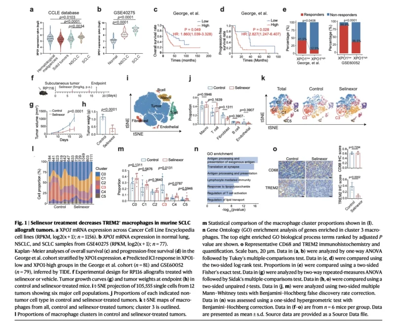
The investigators found XPO1 expression to be elevated in SCLC relative to normal lung, NSCLC and several other tumor contexts. Importantly, expression was distributed across major molecular SCLC subtypes rather than being restricted to a single lineage.
Higher XPO1 expression was associated with worse outcomes in retrospective datasets:
- Overall survival: HR 1.86
- Progression-free survival: HR 2.83
Patients with higher XPO1 expression were also computationally predicted to derive less benefit from immune checkpoint blockade. These associations alone would not establish causality. The more informative experiments came from manipulating XPO1 directly. Treatment of murine SCLC tumors with the XPO1 inhibitor selinexor reduced tumor growth and altered the macrophage compartment.
Single-cell RNA sequencing of 105,555 cells showed that the overall abundance of major immune and stromal populations was not dramatically changed. Instead, the effect was more selective: XPO1 inhibition reduced a macrophage cluster characterized by expression of TREM2 and CD206, together with other immunoregulatory features.
This distinction is important. XPO1 inhibition did not simply remove macrophages from the tumor. It appeared to change the type of macrophages present.
TREM2-Positive Macrophages Emerge as an Immunosuppressive Population
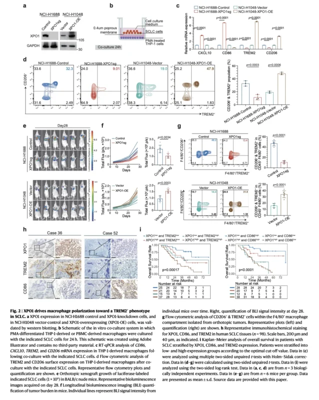
The investigators subsequently demonstrated that tumor-cell XPO1 could actively influence macrophage polarization. When macrophages were co-cultured with SCLC cells in which XPO1 had been depleted, the proportion of CD206+/TREM2+ macrophages fell, while CD86 expression increased.
The opposite occurred when XPO1 was overexpressed. TREM2-positive macrophages also secreted higher levels of immunosuppressive cytokines including IL-10 and TGF-β, while CD8-positive T cells exposed to these macrophages showed reduced IFN-γ expression.
These observations suggest that XPO1 is doing more than supporting tumor-cell survival. It may allow the tumor to actively educate surrounding macrophages toward an immune-suppressive state.
That makes XPO1 particularly interesting therapeutically because inhibition could potentially exert two effects simultaneously, direct tumor-cell pressure, and remodeling of the tumor immune microenvironment.
Human SCLC Samples Support the Biological Association
The investigators also examined 98 surgically resected human SCLC specimens. High tumor XPO1 expression was associated with higher TREM2 expression and lower CD86 expression.
Patients with XPO1-low/TREM2-low or XPO1-low/CD86-high tumors had more favorable overall survival, whereas XPO1-high tumors associated with a TREM2-dominant or CD86-low macrophage phenotype had poorer outcomes.
Multivariable analyses identified the XPO1-high/CD86-low and XPO1-high/TREM2-high phenotypes as independent adverse prognostic factors in that cohort. Again, this does not prove that XPO1 causes poorer survival in patients.
But it strengthens the connection between the mechanistic experiments and human SCLC biology.
The Mechanistic Link: XPO1 Suppresses TNFSF15 Through IRF3
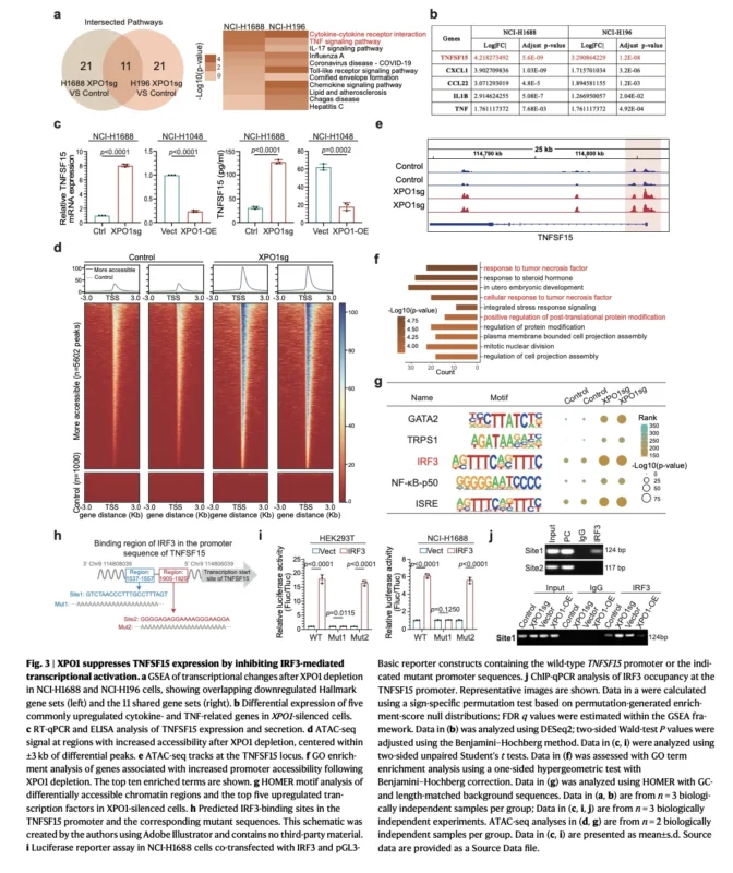
The study then traces the pathway upstream. Following XPO1 depletion, transcriptomic analyses identified TNFSF15 as one of the most strongly increased cytokine-related genes.
Experimental validation showed that XPO1 knockdown increased TNFSF15 expression and secretion, whereas XPO1 overexpression suppressed it. In a human SCLC dataset, XPO1 and TNFSF15 expression were inversely correlated.
The investigators also performed rescue experiments. Blocking TNFSF15 partially reversed the reduction in TREM2-positive macrophages caused by XPO1 loss. Conversely, restoring TNFSF15 counteracted the ability of XPO1 overexpression to push macrophages toward the TREM2-positive phenotype.
TNFSF15 therefore appears to serve as an important tumor-to-macrophage signaling mediator.
But another question remained:
- How does XPO1 suppress TNFSF15?
- The answer appears to involve IRF3.
Chromatin-accessibility and transcription-factor analyses identified increased IRF3 activity around the TNFSF15 promoter after XPO1 loss. Functional experiments then demonstrated that IRF3 can directly promote TNFSF15 transcription.
So the pathway begins to take shape:
- more functional IRF3 → more TNFSF15 → fewer
- immunosuppressive TREM2+ macrophages.
XPO1 interrupts that sequence.
TRIM21 Connects XPO1 to IRF3 Degradation
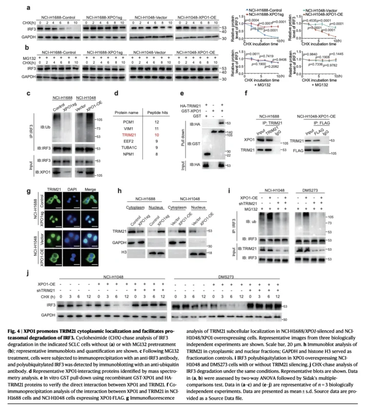
One of the most mechanistically interesting findings concerns TRIM21, an E3 ubiquitin ligase. XPO1 manipulation altered IRF3 protein abundance without significantly changing IRF3 mRNA expression, suggesting post-translational regulation.
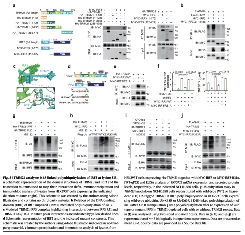
Further experiments demonstrated that XPO1 promoted IRF3 ubiquitination and proteasomal degradation. Mass spectrometry then identified TRIM21 as a candidate mediator. The proposed model is that XPO1 facilitates the nuclear export of TRIM21. Once exported, TRIM21 interacts with IRF3 and promotes its ubiquitination and degradation.
The study further identified K313 as an important IRF3 ubiquitination site in this system. This provides the molecular bridge connecting nuclear transport to immune suppression.
The pathway can therefore be summarized as:
- XPO1 ↑
→ TRIM21 nuclear export ↑
→ IRF3 degradation ↑
→ TNFSF15 ↓
→ TREM2+ macrophage polarization ↑
→ IFN-γ/STAT1 signaling ↓
→ MHC class I ↓
→ CD8+ T-cell recognition ↓
→ immune escape
Blocking XPO1 reverses the cascade.
Restoring MHC Class I May Be the Critical Link to Immunotherapy
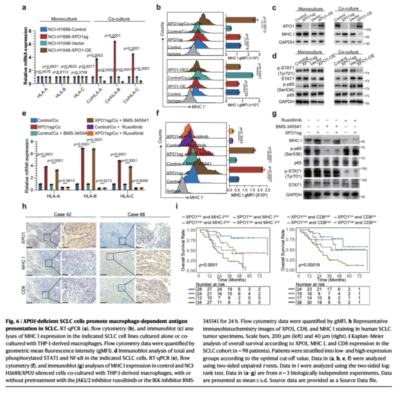
Perhaps the most clinically relevant part of the model involves antigen presentation. XPO1 expression inversely correlated with HLA-A, HLA-B and HLA-C expression in human datasets. Interestingly, manipulating XPO1 in SCLC cells cultured alone did not substantially alter MHC class I.
The effect emerged when tumor cells were co-cultured with macrophages. Under those conditions, XPO1 depletion increased MHC class I expression, while XPO1 overexpression reduced it.
This suggests that XPO1 does not simply regulate MHC I directly within the tumor cell. Instead, it reshapes a communication circuit between the tumor and its macrophage environment.
The investigators traced this effect primarily to IFN-γ–JAK/STAT1 signaling. Blocking JAK1/2, silencing STAT1 or neutralizing IFN-γ attenuated the increase in MHC class I associated with XPO1 loss.
That is potentially important for PD-1 therapy. Checkpoint inhibitors cannot rescue an antitumor T-cell response effectively if tumor cells remain poorly visible to those T cells.
XPO1 inhibition may therefore function less as another immune checkpoint and more as an intervention that makes the tumor immunologically visible again.
The Human Tissue Findings Point in the Same Direction
In the same cohort of 98 resected SCLC specimens, tumors with low XPO1 expression showed higher MHC class I expression, and greater CD8-positive T-cell infiltration. Patients whose tumors combined low XPO1 with high MHC I or high CD8 infiltration also had better overall survival.
This gives the proposed pathway a clinically recognizable phenotype: XPO1-high tumors appear more immune-excluded, while XPO1-low tumors appear more antigen-presenting and T-cell–infiltrated.
The possibility that this phenotype could eventually serve as a biomarker deserves further investigation.
Selinexor Enhanced PD-1 Blockade in Preclinical Models
The translational centerpiece of the study is the combination experiment. Investigators used a humanized orthotopic SCLC model and compared:
- anti-PD-1 alone
- selinexor alone,
- and the combination.
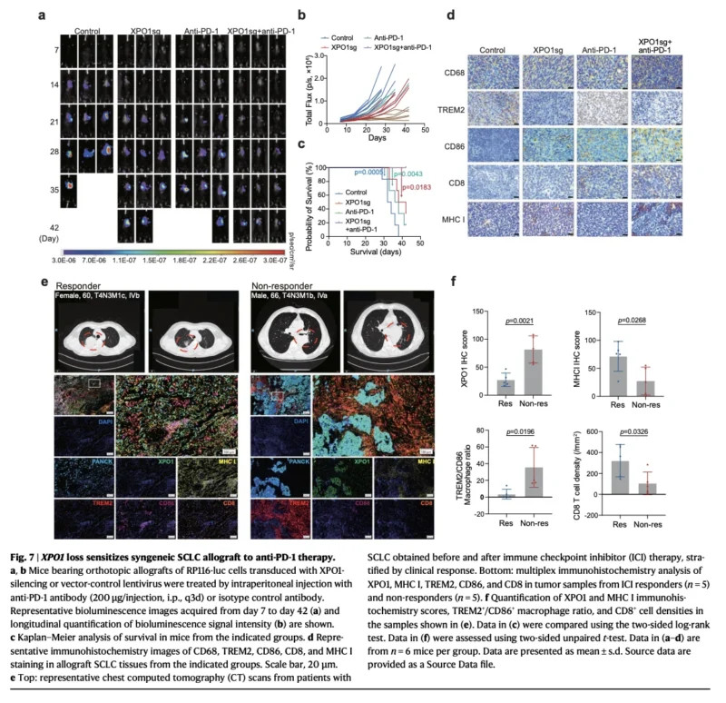
The combination produced greater tumor reduction and longer survival than either monotherapy or control. Tumors treated with combined XPO1 inhibition and PD-1 blockade showed broad immune remodeling more CD86-positive macrophages, fewer TREM2-positive macrophages, higher MHC class I expression and greater CD8-positive T-cell infiltration.
The investigators then reproduced the concept genetically. In immunocompetent mice, XPO1 knockdown combined with anti-PD-1 produced greater tumor suppression and the longest survival among the tested groups.
This is the key therapeutic signal. XPO1 inhibition did not merely show independent antitumor activity. It appeared to sensitize SCLC to PD-1 blockade.
Patient Biopsies Provide an Intriguing Correlative Signal
The investigators also examined tumor biopsies from patients with advanced SCLC who had received first-line immune checkpoint inhibitor therapy.
Responders had:
- lower XPO1 expression (P=.0021),
- higher MHC class I expression (P=.0268),
- a lower TREM2+/CD86+ macrophage ratio (P=.0196),
- and greater CD8+ T-cell infiltration (P=.0326)
than nonresponders.
Responders also had CD8-positive T cells located closer to tumor cells. These findings are compelling because they align remarkably well with the proposed experimental mechanism. But they remain correlative. They do not demonstrate that giving an XPO1 inhibitor would convert an ICI nonresponder into a responder.
That is the therapeutic hypothesis that now needs prospective testing.
This Is Not Yet a New SCLC Treatment Strategy
The translational enthusiasm around these findings should be matched by equally careful restraint.
No patients in this study were prospectively treated with selinexor plus anti-PD-1 therapy to determine whether the combination improves response rate, PFS or overall survival. The therapeutic evidence comes from mouse models.
The human data establish associations between XPO1, macrophage state, antigen presentation, T-cell infiltration, prognosis and ICI response, but not clinical efficacy of XPO1 inhibition.
The authors themselves identify several unresolved questions. XPO1 may influence T cells through mechanisms not fully characterized. Its effects on macrophage phagocytosis and migration remain uncertain. The precise mechanism through which macrophages regulate STAT1 signaling in tumor cells requires further clarification.
And although TNFSF15 appears important, other cytokines altered after XPO1 loss, including CXCL1, CCL22 and IL1B, may contribute to the phenotype. SCLC heterogeneity is another important consideration.
The human cohorts are relatively limited, and differences in molecular subtype, treatment history and cohort composition could affect generalizability.
Even the proposed mechanism of TRIM21 nuclear export will require additional validation, including experiments specifically interrogating the nuclear export signal and alternative cofactors.
The More Important Concept May Be Macrophage Reprogramming
This paper is important not simply because it identifies another potential SCLC drug combination. Its broader contribution is to change how immune resistance is conceptualized. Checkpoint resistance is often framed primarily as a T-cell problem. But the immune system around a tumor is an ecosystem.
A PD-1 antibody cannot generate effective tumor control if tumor cells remain poorly visible, antigen presentation is suppressed and macrophages maintain an immunoregulatory environment hostile to productive T-cell activity.
The findings suggest that TREM2-positive macrophages may represent one component of that barrier in SCLC. Rather than eliminating macrophages indiscriminately, XPO1 blockade appeared to shift macrophage state while leaving total macrophage abundance relatively unchanged.
That may be a particularly attractive therapeutic concept. The objective is not necessarily to remove the myeloid compartment. It may be to reprogram it.
From Immune Checkpoint Blockade to Immune-Ecosystem Repair
The current immunotherapy paradigm in SCLC largely focuses on releasing inhibitory signals from T cells. This study suggests a complementary strategy.
Before asking a T cell to attack, perhaps the tumor microenvironment must first be made capable of presenting antigen and supporting immune recognition.
In the proposed model:
- XPO1 inhibition restores IRF3
- IRF3 restores TNFSF15
- TNFSF15 constrains TREM2-positive macrophage polarization
- macrophage remodeling restores IFN-γ/STAT1 signaling
- MHC class I increases
- CD8-positive T cells gain greater access to an immunologically visible tumor
Only then may PD-1 blockade have a more favorable environment in which to operate. That is why the study is conceptually interesting. It frames XPO1 inhibition not simply as direct anticancer therapy, but as a potential way to repair the immune context in which checkpoint blockade is being asked to work.
The Bottom Line
This Nature Communications study identifies XPO1 as a potential regulator of immune exclusion in small cell lung cancer. The investigators propose a mechanistic cascade in which XPO1-dependent TRIM21 nuclear export promotes IRF3 degradation, suppresses TNFSF15 and favors accumulation of immunosuppressive TREM2-positive macrophages.
The downstream consequence is reduced macrophage-dependent IFN-γ/STAT1 activity, diminished MHC class I expression and impaired CD8-positive T-cell recruitment.
Genetic or pharmacological XPO1 inhibition reversed these effects and enhanced anti-PD-1 activity in preclinical SCLC models. Human tissue analyses showed that lower XPO1 expression was associated with higher MHC I, greater CD8 infiltration and more favorable clinical features.
The translational hypothesis is therefore compelling:
Could XPO1 inhibition convert an immune-resistant SCLC microenvironment into one that is more responsive to checkpoint blockade?
The current study provides mechanistic support for asking that question. It does not yet provide the clinical answer. Prospective trials will need to establish whether the biological reprogramming observed in these models can translate into meaningful improvements in response, survival and tolerability for patients with SCLC.
Reference
- Du Q, Qin T, Wang J, et al. XPO1-mediated TRIM21 nuclear export reprograms TREM2+ macrophage polarization by targeting IRF3 to augment anti-PD-1 efficacy in small cell lung cancer. Nature Communications. Article in Press. Accepted August 6, 2026. doi:10.1038/s41467-026-76862-0.