Thoracic radiotherapy has become increasingly precise.
Modern treatment planning carefully limits radiation exposure to the lungs, heart, esophagus, spinal cord and other structures where dose can translate into clinically meaningful toxicity.
But one organ has largely remained outside this conversation: the thymus.
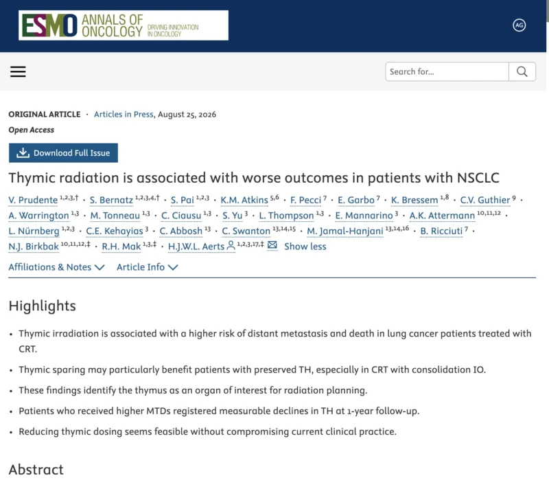
A new multicohort analysis published in Annals of Oncology suggests that this omission may matter.
Across 1,107 patients with locally advanced non small-cell lung cancer, higher incidental radiation exposure to the thymus was associated with a greater risk of distant metastasis and, in several analyses, worse overall survival. The association appeared particularly pronounced in patients who entered treatment with relatively preserved thymic function.
The findings are not sufficient to establish thymic dose constraints or immediately redefine radiotherapy planning.
But they raise a provocative clinical question:
When we irradiate locally advanced lung cancer, could we also be damaging an organ that contributes to the systemic immune response needed to prevent metastatic relapse?
That question becomes especially important in the modern era of chemoradiotherapy followed by immune checkpoint inhibition.
The Adult Thymus May Matter More Than We Thought
The thymus is central to T-cell development, but its importance has traditionally been associated mainly with childhood.
With aging, functional thymic tissue is progressively replaced by fat, leading to the longstanding assumption that the adult thymus has limited clinical relevance.
That view is changing.
Recent work has suggested that residual thymic function persists into adulthood and may influence immune competence, cancer outcomes and response to immunotherapy. The investigators behind the current study had previously developed a deep-learning imaging biomarker called Thymic Health, or TH, designed to estimate thymic functional status from routine thoracic CT imaging.
That creates a new way to think about thoracic radiotherapy.
The thymus lies in the anterior mediastinum, close enough to many thoracic radiation fields to receive substantial incidental exposure. Yet unlike the heart, lungs or esophagus, it is not routinely delineated or treated as an organ at risk.
The biological concern is straightforward.
Radiotherapy can produce immunogenic tumor-cell death and potentially enhance antitumor immune responses. At the same time, radiation can also damage lymphocytes and immune structures.
In an era when the success of treatment increasingly depends on the interaction between radiotherapy and immunotherapy, preserving immune competence may become part of optimizing local therapy.
Three Cohorts, More Than 1,100 Patients
Prudente and colleagues evaluated 1,107 patients with locally advanced NSCLC across three independent cohorts.
The analysis included 460 patients from the prospective Phase 3 RTOG-0617 trial, 422 real-world patients treated with chemoradiotherapy without consolidation immunotherapy, and 225 patients treated with chemoradiotherapy followed by durvalumab.
Using an automated deep-learning segmentation system, the investigators identified the thymic bed on treatment-planning CT scans and calculated radiation exposure, including the mean thymic dose, or MTD.
They separately estimated baseline thymic function using the previously developed TH imaging model, which assigns a value from 0 to 1 based on radiographic characteristics of the thymus. Patients were classified as having preserved or impaired thymic health.
The primary clinical endpoint was distant metastasis.
That choice is important.
If thymic irradiation simply produced another form of local thoracic toxicity, one might expect its main impact to appear through morbidity or treatment tolerance.
Instead, the investigators were testing whether injury to an immune organ might influence systemic tumor control.
Higher Thymic Dose Was Associated With More Distant Metastasis
The signal appeared consistently across the three cohorts.
In RTOG-0617, increasing mean thymic dose remained associated with distant metastasis after adjustment for baseline thymic health, heart and lung radiation exposure, stage, smoking history and planning target volume.
The authors estimated that each additional 1 Gy of thymic exposure corresponded to approximately a 1.57% increase in distant-metastasis risk. In the adjusted model, the hazard ratio was 1.29 per standard-deviation increase in MTD. Higher MTD was also associated with increased mortality, with an adjusted HR of 1.25.
The association was reproduced in the independent HARVARD-CRT cohort.
There, each additional 1 Gy was associated with an estimated 1.76% increase in distant-metastasis risk, with an adjusted HR of 1.33 per standard-deviation increase in MTD.
The most striking signal emerged in patients receiving contemporary treatment with consolidation durvalumab.
Among the HARVARD-DURVA cohort, each additional 1 Gy in mean thymic dose was associated with an estimated 4.21% increase in the risk of distant metastasis among patients with preserved baseline thymic health. The corresponding adjusted HR was 1.95 per standard-deviation increase in thymic dose.
That observation is particularly interesting because this is precisely the clinical setting in which treatment success depends partly on sustained antitumor immunity after chemoradiation.
The 35-Gy Signal Is Interesting, but Not a Clinical Constraint
The investigators also explored whether a specific thymic dose might separate higher- and lower-risk patients.
They selected a mean thymic dose of 35 Gy from the RTOG-0617 dataset as an exploratory threshold.
In RTOG-0617, patients receiving more than 35 Gy to the thymus had a 40% higher unadjusted risk of distant metastasis than those receiving lower doses.
The same threshold was associated with a 57% greater risk in the HARVARD-CRT cohort and nearly twice the risk of distant metastasis in the HARVARD-DURVA cohort.
Those numbers are provocative.
They should not, however, be converted into a new radiotherapy planning rule.
The authors explicitly emphasize that the 35-Gy cut-off was exploratory, population-specific and not prospectively validated. The underlying dose-response analysis did not identify one clear biological threshold; rather, a range between approximately 25 and 50 Gy appeared potentially relevant.
So the current message is not:
- Keep the thymus below 35 Gy.
The more appropriate conclusion is:
Thymic dose may matter, and prospective work is needed to determine whether a clinically meaningful constraint exists.
That distinction is essential.
Baseline Thymic Health May Identify Who Is Most Vulnerable
One of the most biologically interesting findings was that radiation exposure did not appear to carry the same implications for every patient.
The adverse association between thymic dose and clinical outcome was strongest among patients who began treatment with preserved thymic health.
At first glance, that might seem paradoxical.
Why would a healthier thymus identify a patient at greater risk?
The likely explanation is that there is more functional immune capacity to lose. If the thymus is already severely involuted or dysfunctional, additional radiation injury may have comparatively little measurable effect.
In contrast, a patient with preserved thymic function may lose an active source of immune competence when that organ receives substantial radiation.
This interaction was especially apparent in the durvalumab-treated cohort: increasing MTD was associated with distant-metastasis risk among patients with preserved TH, while essentially no corresponding effect was seen in patients with impaired TH.
The implication is potentially important. A future thymus-sparing strategy might not need to be applied identically to every patient. Instead, baseline imaging or other measures of thymic function could eventually identify individuals most likely to benefit from immune-organ preservation.
That would convert the concept from simple anatomical sparing toward biologically personalized radiotherapy.
Radiation Appeared to Damage the Thymus Over Time
Association with clinical outcome alone would still leave open the possibility that thymic dose was simply a surrogate for more difficult disease geometry. The investigators therefore looked for evidence that radiation exposure actually changed thymic health.
Among 209 patients with baseline and approximately 1-year follow-up imaging, patients receiving at least 35 Gy demonstrated a significant decline in TH, whereas those below that threshold did not.
Higher mean thymic dose was independently associated with lower TH at follow-up. A much smaller subset of 40 patients also had longitudinal lymphocyte measurements.
Patients with higher thymic exposure tended to have lower circulating lymphocyte counts at follow-up, although the difference narrowly missed statistical significance.
These analyses remain exploratory, but they strengthen the biological plausibility of the clinical observation.
The proposed sequence is:
thymic irradiation → thymic functional decline → impaired T-cell competence → weaker systemic tumor surveillance → increased metastatic risk.
The study does not prove that causal pathway.
But it provides enough concordant evidence to justify testing it prospectively.
Could Thymic Injury Affect the Interaction Between Radiation and Immunotherapy?
This may be the most consequential question raised by the study.
Chemoradiotherapy followed by durvalumab transformed the treatment of unresectable stage III NSCLC.
The therapeutic model implicitly relies on two treatment effects working together: radiation provides local control and antigen release, while immune checkpoint inhibition sustains or amplifies systemic antitumor immunity.
But what happens if radiation simultaneously injures an organ involved in maintaining the T-cell repertoire?
The authors suggest that this could help explain why some attempts to intensify or combine thoracic radiotherapy and immunotherapy have not translated into better outcomes.
That remains hypothesis-generating rather than proven.
Still, the biological tension is compelling:
radiotherapy may stimulate antitumor immunity while simultaneously damaging the infrastructure required to sustain it.
The timing, volume and dose of radiation to immune structures could therefore become relevant not only to toxicity, but also to the effectiveness of immunotherapy.
The study authors specifically note that the association between thymic dose and outcomes was seen in both pre-PACIFIC cohorts and in patients treated with consolidation durvalumab, although they appropriately caution that treatment heterogeneity and selection bias limit cross-cohort interpretation.
Can the Thymus Actually Be Spared?
A biomarker matters clinically only if something can be done about it.
The investigators therefore performed an exploratory replanning analysis in 16 patients treated with modern radiotherapy techniques.
The original treatment plans were reoptimized with thymic dose reduction incorporated into the planning objectives.
Thymic exposure fell significantly, without a meaningful deterioration in target coverage or increased radiation exposure to other major organs at risk.
The dosimetric analysis on page 10 is particularly relevant: the replanned treatments produced substantial reductions in thymic dose while preserving planning-target-volume coverage and maintaining lung, esophageal and spinal-canal constraints.
This does not establish clinical benefit.
Sixteen replanned cases are a feasibility experiment, not a treatment trial.
But the finding is important because it suggests that the hypothesis may be testable without sacrificing established oncologic priorities.
The next step is therefore technically conceivable: prospectively compare immune and oncologic outcomes in patients treated with standard versus thymus-conscious planning.
Why This Study Should Not Yet Change Routine Practice
The study has several important limitations.
Although one dataset came from the prospective RTOG-0617 trial, the thymic-dose analysis itself was not based on randomization to thymic exposure.
The two Harvard cohorts were retrospective.
There was no prospective thymus-sparing intervention, no randomized comparison according to the 35-Gy threshold, and no evidence yet that deliberately lowering thymic dose reduces metastatic recurrence.
The population was predominantly White, and the deep-learning TH model requires broader validation across institutions, scanners and populations.
Biological validation was also limited: longitudinal lymphocyte data were available in only 40 patients, while key factors including PD-L1, tumor mutational burden, driver mutations, comorbidity and some inflammatory biomarkers were unavailable or incomplete.
Residual confounding therefore remains possible.
These limitations matter because treatment geometry can itself reflect disease biology.
A tumor requiring broader or more central radiation fields may differ clinically from one allowing easier thymic avoidance, even if statistical models adjust for stage, target volume and doses to surrounding organs.
The association is therefore compelling, reproducible and biologically plausible, but still associative rather than causal.
A New Definition of an Organ at Risk?
The concept of an organ at risk has traditionally focused on preventing structural toxicity. We protect the lungs to reduce pneumonitis. We protect the heart to reduce cardiovascular disease.
We protect the spinal cord because exceeding tolerance can produce catastrophic neurological injury. The thymus introduces a different concept.
Its potential toxicity may not manifest primarily as damage to the organ itself. Instead, the consequence could be loss of systemic immune competence. That means future radiation planning may eventually have to distinguish between two categories of protected structures:
organs whose injury causes direct local toxicity, and organs whose injury compromises the patient’s ability to control cancer systemically. The spleen, bone marrow, circulating lymphocyte pool and now potentially the thymus all fit into this broader concept of an immune organ at risk.
That could become particularly important as radiotherapy is combined with immunotherapy across more tumor types and disease stages.
The Bottom Line
This study does not establish a new thymic radiation-dose constraint.
It establishes a reason to investigate one.
Across three independent NSCLC cohorts comprising 1,107 patients, higher incidental thymic radiation exposure was consistently associated with increased distant-metastasis risk, with the strongest signal in patients who had preserved thymic function before treatment.
Higher thymic dose was also associated with subsequent decline in imaging-defined thymic health, providing a potential biological link between radiation exposure and impaired immune function.
Most importantly, exploratory replanning suggests that thymic exposure can potentially be reduced without compromising tumor coverage or established organ-at-risk constraints.
The findings are therefore hypothesis-generating, not practice-defining.
But they introduce an important new question into thoracic oncology:
Should successful radiotherapy mean not only controlling the tumor and protecting the heart and lungs, but also preserving the immune system that must control disease after radiation ends?
If prospective studies confirm that thymic sparing reduces metastatic relapse, particularly in patients receiving immunotherapy, the thymus could move from an almost ignored mediastinal structure to a meaningful component of personalized thoracic radiotherapy planning.
That would represent a larger shift than simply adding one more contour.
It would mean treating immune health itself as an organ at risk.
Reference
- Prudente V, Bernatz S, Pai S, et al. Thymic radiation is associated with worse outcomes in patients with NSCLC. Annals of Oncology. 2026. doi:10.1016/j.annonc.2026.07.001.