A new real-world study has identified a potentially underrecognized pattern of acute neurological toxicity in patients with extensive-stage small cell lung cancer (ES-SCLC) receiving tarlatamab. Investigators from the University Hospital Cologne reported neurological deterioration consistent with tumor inflammation-associated neurotoxicity (TIAN) in seven of 41 treated patients, all of whom had pre-existing brain metastases. The findings suggest that some neurological events occurring shortly after tarlatamab administration may involve localized inflammatory changes rather than classical immune effector cell-associated neurotoxicity syndrome (ICANS).
The findings, reported by Malte Verheyen and colleagues in an accepted article for JTO Clinical and Research Reports, raise important questions about neurological assessment, adverse-event classification, and the management of patients with central nervous system (CNS) involvement.
Among the seven suspected TIAN cases, six patients experienced improvement after dexamethasone, with a median reported time to improvement of 20.5 hours. The remaining patient’s symptoms resolved spontaneously within 72 hours.
A particularly important observation was that neurological deterioration sometimes occurred despite normal or only mildly impaired Immune Effector Cell-Associated Encephalopathy (ICE) scores. This suggests that conventional ICANS-oriented assessment may not adequately capture focal neurological complications in patients with brain metastases.
However, the study was retrospective, involved a small number of patients, and relied on clinical classification rather than prospectively validated diagnostic criteria. The findings should therefore be considered an important safety signal requiring further investigation, not proof that TIAN represents a fully established independent toxicity syndrome.
Tarlatamab Expands Treatment Opportunities in Small Cell Lung Cancer
Small cell lung cancer remains an aggressive malignancy characterized by rapid progression, frequent early dissemination, and a high incidence of brain metastases. Although platinum-based chemotherapy combined with immunotherapy has improved outcomes in extensive-stage disease, treatment options after progression have historically offered limited durable benefit.
Tarlatamab represents a major therapeutic advance in this setting. The agent is a DLL3-directed bispecific T-cell engager designed to simultaneously bind delta-like ligand 3 (DLL3) on SCLC cells and CD3 on T lymphocytes. This interaction promotes immune-cell recruitment and T-cell-mediated destruction of tumor cells.
Clinical studies, including DeLLphi-301 and the randomized phase III DeLLphi-304 trial, established substantial antitumor activity and improved outcomes compared with conventional chemotherapy in previously treated SCLC.
These findings supported the integration of tarlatamab into the treatment landscape for patients progressing after platinum-based chemotherapy. However, T-cell-engaging therapies introduce distinct immune-related toxicities.
The best characterized are cytokine release syndrome (CRS) and ICANS, both of which may require treatment interruption, corticosteroids, supportive care, and intensive monitoring depending on severity.
As tarlatamab use expands into routine clinical practice and earlier treatment settings, understanding the full spectrum of neurological adverse events has become increasingly important.
Why TIAN May Represent a Different Neurological Complication
Tumor inflammation-associated neurotoxicity was initially described in patients receiving chimeric antigen receptor T-cell therapies, particularly when tumors were located within the CNS. The proposed mechanism involves localized inflammation in or around tumor deposits following immune activation.
Unlike classical ICANS, which frequently produces diffuse neurological dysfunction, TIAN may manifest through focal symptoms determined by the anatomical location of pre-existing CNS lesions. These can include worsening hemiparesis, paresthesias, headaches, altered motor function, or other neurological deficits associated with localized edema.
Such symptoms may resemble tumor progression, radiation-related changes, intracranial hypertension, or conventional ICANS. Differentiating these processes is clinically important because their evaluation and management may differ.
A previous case report had described TIAN-like neurological deterioration after tarlatamab treatment, but systematic evidence remained limited. The Cologne investigators therefore examined their institutional experience to determine whether similar presentations could be identified in a consecutive real-world cohort.
Study Design: Real-World Neurological Safety Assessment
The retrospective single-center study included 41 patients with ES-SCLC who received at least one dose of tarlatamab at the University Hospital Cologne between May 8, 2025, and March 31, 2026. Patients received tarlatamab using a step-up dosing schedule, beginning with a 1-mg dose followed by escalation to 10 mg.
All patients were treated as inpatients and monitored for at least 48 hours following each of the first two administrations under the institution’s protocol. Clinical monitoring included repeated assessments of vital signs, pain severity, neurological symptoms, and cognitive function using the ICE score.
Neurological events were identified retrospectively from medical records and assessed using CTCAE Version 6.0 terminology. Two experienced thoracic oncologists independently reviewed suspected events, with disagreements resolved through consultation with a third senior physician.
The analysis aimed to distinguish clinical presentations consistent with TIAN from ICANS and other possible causes of neurological deterioration. Importantly, the investigators did not use a prospectively validated diagnostic test for TIAN. Classification was based on clinical presentation, temporal relationships, available neuroimaging, and response to treatment.
TIAN-Like Symptoms Were Identified in Seven Patients
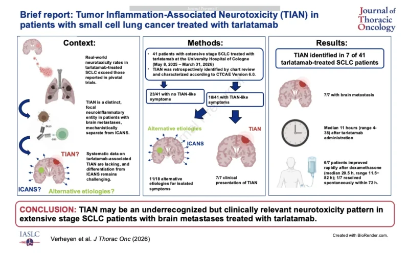
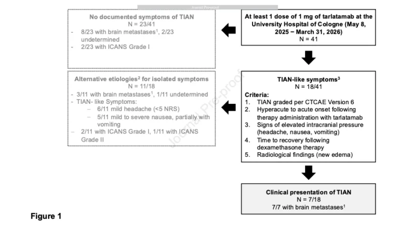
Among the 41 patients treated with tarlatamab, 18 developed symptoms potentially consistent with TIAN. After clinical review, 11 of these cases were attributed to alternative explanations, including nonspecific headache, nausea, and other symptoms.
The remaining seven patients, representing approximately 17% of the entire cohort, were retrospectively classified as having neurological deterioration clinically consistent with TIAN. All seven had documented brain metastases before starting tarlatamab.
This was one of the most notable findings. The observations suggest that pre-existing CNS tumor involvement may be an important characteristic of patients developing this neurological phenotype. However, the study does not establish the incidence of TIAN among all patients with brain metastases, nor does it prove that brain metastases are an independent predictive risk factor.
The study population was small, the number of suspected events was limited, and other patient or treatment characteristics could not be adequately evaluated. Nevertheless, the consistent presence of brain metastases among affected patients provides a biologically plausible basis for further investigation.
Neurological Deterioration Occurred Within Hours of Treatment
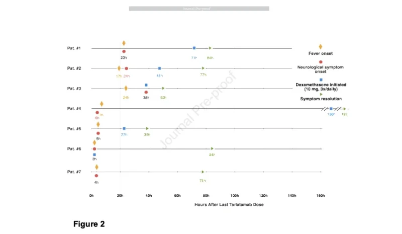
The suspected TIAN cases were characterized by rapid symptom onset following tarlatamab administration. Most events occurred after the initial 1-mg priming dose, while two occurred following the subsequent 10-mg dose.
Symptoms included acute headache, dizziness, confusion, somnolence, worsening paresthesias, and deterioration of existing motor deficits. The clinical presentations were heterogeneous, reflecting the different locations and burdens of intracranial disease.
One patient with a pre-existing thalamic metastasis developed worsening hemiparesis following the second tarlatamab administration. Subsequent imaging demonstrated increased edema around the known lesion with compression of the third ventricle, consistent with an inflammatory or pseudoprogression-like process.
Another patient with previously stable brain metastases developed severe headache, dizziness, and psychomotor slowing soon after the first dose. Other patients experienced new or worsening neurological symptoms despite no immediately documented radiographic progression.
The rapid timing of these events may help distinguish a treatment-associated inflammatory process from some forms of conventional ICANS. However, timing alone is insufficient for diagnosis, and early ICANS, intracranial disease progression, seizures, vascular events, and other neurological complications must remain in the differential diagnosis.
ICE Scores May Fail to Identify Focal Neurological Toxicity
Perhaps the most important clinical finding concerns the limitations of conventional cognitive assessment. The ICE score is commonly used to evaluate immune effector cell-associated encephalopathy, including orientation, naming, writing, attention, and command-following abilities.
However, it was designed primarily to identify global cognitive dysfunction. It does not directly quantify focal motor weakness, localized sensory abnormalities, cranial nerve dysfunction, or other regionally restricted neurological changes.
In the Cologne cohort, several patients experienced clinically important neurological symptoms while their ICE scores remained normal or only mildly abnormal. Two patients retained ICE scores of 10/10 despite neurological deterioration.
Others had scores of 8/10 or 9/10 in association with headaches, dizziness, or focal deficits. One patient experienced more severe neurological dysfunction, with the ICE score decreasing to 3/10, raising the possibility of overlapping TIAN and ICANS manifestations.
These findings suggest that an ICE score alone may not be sufficient to exclude clinically significant neurological toxicity. A patient with a normal score may still have worsening weakness, new paresthesias, severe headache, or evolving edema around intracranial metastases.
The authors therefore emphasize the importance of comprehensive baseline neurological examinations and repeated focused assessments during tarlatamab treatment. This is particularly relevant for patients with known brain metastases or pre-existing neurological deficits.
Dexamethasone Was Associated With Rapid Symptom Improvement
Six of the seven patients classified as having suspected TIAN received dexamethasone. Symptom improvement occurred after a median of 20.5 hours, with a reported range of 11.5–82 hours.
The remaining patient did not receive corticosteroids, and symptoms resolved spontaneously within 72 hours. In several cases, corticosteroid administration was followed by rapid improvement in headache, focal weakness, or cognitive symptoms.
The investigators interpreted these responses as consistent with an inflammatory component. However, corticosteroid responsiveness is not specific to TIAN. Dexamethasone can also improve symptoms caused by peritumoral edema, increased intracranial pressure, and other inflammatory neurological processes.
Consequently, the observed improvement supports the plausibility of localized inflammation but does not independently establish the diagnosis. The case series also cannot determine the optimal corticosteroid regimen, timing, or duration.
The authors suggest that early treatment may be appropriate when a clinically concerning TIAN-like presentation is suspected, including in patients whose ICE scores remain relatively preserved. This represents a hypothesis-generating recommendation rather than a validated treatment algorithm.
In clinical practice, new or worsening neurological symptoms during tarlatamab treatment require urgent evaluation, consideration of treatment interruption, appropriate imaging, and assessment for ICANS and other potentially life-threatening causes.
Neuroimaging Suggests a Localized Inflammatory Process
Neuroimaging findings provided additional support for the proposed TIAN phenotype in selected cases. In one patient, cerebral imaging demonstrated increasing edema around a known thalamic metastasis, producing greater mass effect and ventricular compression.
Another patient demonstrated edema adjacent to a previously treated intracranial lesion. These findings are compatible with an inflammatory or pseudoprogression-like reaction following immune activation.
However, most patients did not undergo immediate neuroimaging at the time of symptom onset. This represents a substantial limitation because radiological confirmation was unavailable for the majority of suspected cases.
Furthermore, imaging changes around brain metastases can arise from several processes, including tumor progression, prior radiotherapy, edema, hemorrhage, and treatment-associated inflammation. The findings therefore support consideration of TIAN but do not establish a definitive imaging signature.
Prospective studies incorporating standardized baseline MRI, early imaging during symptoms, and longitudinal follow-up will be required to characterize the radiographic manifestations more accurately.
TIAN Versus ICANS: Why the Distinction Matters
The potential distinction between TIAN and ICANS has direct implications for neurological monitoring. ICANS can involve confusion, encephalopathy, impaired attention, language disturbance, seizures, and reduced consciousness.
TIAN, as proposed in this study, may be more closely associated with rapid-onset focal neurological deterioration and localized peritumoral inflammation. Nevertheless, the two syndromes are not necessarily mutually exclusive.
Some patients may demonstrate overlapping features, particularly when localized CNS inflammation is accompanied by more generalized neurological dysfunction. The Cologne investigators described one patient with a presentation suggesting such overlap.
They also noted that applying conventional ICANS grading alone may lead clinicians to underestimate focal neurological deterioration. For example, patients with preserved cognition may have low-grade ICANS scores despite clinically significant neurological deficits.
However, the evidence is not sufficient to establish an entirely separate standardized grading system. Until prospective diagnostic criteria are available, the most practical approach is to assess neurological symptoms comprehensively rather than relying exclusively on a single cognitive score.
Clinical Implications for Patients With Brain Metastases
The findings are particularly relevant because brain metastases are common during the course of ES-SCLC. As effective systemic treatments achieve improved disease control, more patients with active, previously treated, or stable intracranial disease may become candidates for DLL3-directed immunotherapy.
The case series suggests that several clinical precautions deserve attention. Baseline neurological assessment should document pre-existing weakness, sensory abnormalities, cognitive changes, seizures, headaches, and other symptoms.
During the early dosing period, clinicians should remain alert for new focal neurological deficits or deterioration of established symptoms, even when ICE scores are reassuring. Urgent evaluation is appropriate when new or worsening neurological symptoms occur, particularly severe headache, altered consciousness, seizures, or focal weakness.
Assessment may include neurological consultation and brain CT or MRI as clinically indicated, together with consideration of disease progression, edema, hemorrhage, infection, metabolic abnormalities, CRS, and ICANS.
The updated US prescribing information for tarlatamab includes boxed warnings for CRS and neurological toxicity, including ICANS, and provides recommendations for treatment interruption and toxicity management.
The Cologne study should therefore be interpreted as a potential refinement of neurological safety assessment, not as a replacement for established prescribing-information requirements. It also does not support withholding effective tarlatamab treatment from all patients with brain metastases.
Rather, it highlights a population in whom careful clinical monitoring and prompt investigation of neurological symptoms may be especially important.
Limitations and Outstanding Questions
Several limitations prevent definitive conclusions. The study involved only 41 patients at a single institution, with seven events classified as TIAN. Its retrospective design created potential limitations in documentation, symptom identification, and adjudication.
The absence of prospective diagnostic criteria creates uncertainty in distinguishing TIAN from ICANS and other neurological complications. Neuroimaging was unavailable at symptom onset in most patients, limiting confirmation of localized inflammatory changes.
In addition, the investigators could not definitively exclude tumor progression in some cases. The association between TIAN-like symptoms and brain metastases is intriguing but does not establish causality or quantify the risk associated with specific intracranial disease characteristics.
The study also cannot determine whether lesion location, tumor burden, previous cranial radiotherapy, steroid exposure, or neurological symptoms at baseline influence the likelihood of toxicity. Finally, the observed improvement after dexamethasone does not establish superiority over alternative approaches or prove that early corticosteroid treatment should be applied universally.
Prospective multicenter studies incorporating systematic neurological examinations, standardized imaging, and validated toxicity classification will be essential.
OncoDaily Takeaway
This real-world case series identifies a potentially important neurological safety signal in patients with extensive-stage SCLC receiving tarlatamab, particularly those with pre-existing brain metastases. Seven of 41 treated patients experienced neurological deterioration clinically consistent with tumor inflammation-associated neurotoxicity, and all seven had intracranial disease before treatment.
Symptoms generally developed rapidly after tarlatamab administration and included headaches, focal neurological deterioration, dizziness, and altered cognition. Six patients improved following dexamethasone, supporting the possibility of a reversible inflammatory component.
The most important practical finding is that clinically significant neurological deterioration may occur despite normal or only mildly impaired ICE scores. However, TIAN remains an incompletely characterized toxicity pattern, and the small retrospective series does not establish a validated diagnostic or treatment standard.
As tarlatamab becomes more widely integrated into SCLC treatment, these observations reinforce the importance of comprehensive neurological assessment, particularly during early dosing in patients with pre-existing CNS involvement.
The emerging clinical priority is not simply monitoring for ICANS, but recognizing the full spectrum of neurological complications associated with T-cell-engaging therapies—without delaying appropriate investigation or treatment of potentially serious CNS events.
Source
- Verheyen M, Balke-Want H, Eckhoff J, et al. Brief Report: Tumor Inflammation-Associated Neurotoxicity (TIAN) in Patients With Small Cell Lung Cancer Treated With Tarlatamab. JTO Clinical and Research Reports. 2026.