Tarlatamab has expanded treatment options for patients with previously treated small-cell lung cancer. However, its use in patients with active or symptomatic brain metastases remains clinically difficult.
Neurotoxicity is a recognized risk of T-cell-engaging therapies. In a patient with intracranial disease, severe neurologic symptoms after treatment may be attributed to immune effector cell-associated neurotoxicity syndrome, known as ICANS. This diagnosis can lead to permanent discontinuation of treatment, particularly when toxicity meets grade 4 criteria.
A recent case report raises an important alternative explanation.
The report describes a patient with relapsed SCLC and multiple active brain metastases who developed loss of consciousness and seizures 27 hours after her first dose of tarlatamab. Although the event initially met criteria for grade 4 neurotoxicity, the imaging, electroencephalography, clinical recovery, and later tumor response suggested localized tumor inflammation-associated neurotoxicity, or TIAN, rather than classic diffuse ICANS.
The distinction remains investigational. However, it may become increasingly relevant as DLL3-targeted T-cell-engaging therapies are used in patients with CNS involvement.
Why Brain Metastases Remain a Difficult Setting for Tarlatamab
Tarlatamab is a DLL3-targeting bispecific T-cell engager designed to direct cytotoxic T cells toward DLL3-expressing SCLC cells.
The DeLLphi clinical trials established activity for tarlatamab in previously treated SCLC. However, patients with symptomatic or untreated brain metastases were generally excluded. Patients with stable, treated CNS disease could be included, leaving limited evidence for patients with active intracranial tumor burden.
This matters because brain metastases are common in SCLC and often progress despite radiotherapy, corticosteroids, antiseizure medications, and systemic treatment.
When neurologic symptoms occur after a T-cell engager in this setting, several possibilities must be considered: ICANS, seizure activity, tumor progression, pseudoprogression, hemorrhage, infection, metabolic encephalopathy, increased intracranial pressure, or treatment-related inflammation around brain metastases.
A Young Patient With Active CNS Disease
The report involved a 26-year-old woman with stage IVB SCLC and metastatic disease involving the liver, pancreas, brain, and spinal cord.
She had previously received cisplatin, etoposide, and durvalumab, followed by whole-brain radiotherapy, surgical debulking of a symptomatic left temporal brain metastasis, and irinotecan.
Before tarlatamab treatment, she had more than 10 active brain metastases involving the frontal, temporal, and parietal lobes. The largest lesion was approximately 30 mm. She also had spinal cord metastases.
The patient had a prior history of tumor-related epilepsy. Her seizures had been controlled with levetiracetam for more than two months before tarlatamab was initiated.
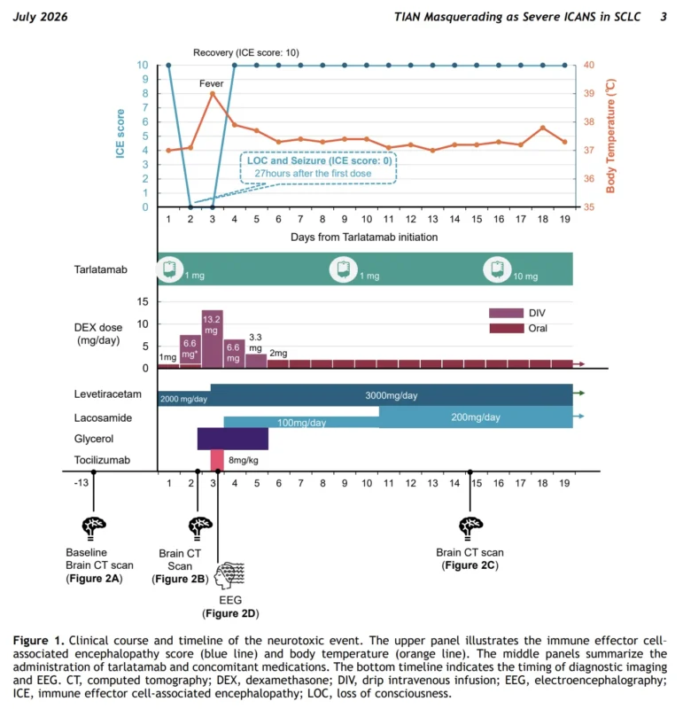
Tarlatamab was started as fourth-line therapy at a 1 mg dose, with dexamethasone prophylaxis and continuation of maintenance corticosteroids and levetiracetam.
Sudden Seizures and Loss of Consciousness After the First Dose
Approximately 24 hours after the first infusion, the patient developed nausea and vomiting.
Three hours later, she experienced right-sided gaze deviation followed by focal-to-bilateral tonic-clonic seizures, loss of consciousness, and an immune effector cell-associated encephalopathy score of 0.
The event occurred 27 hours after tarlatamab administration.
Her Glasgow Coma Scale score was 3. Under conventional grading criteria, the presentation was consistent with grade 4 neurotoxicity.
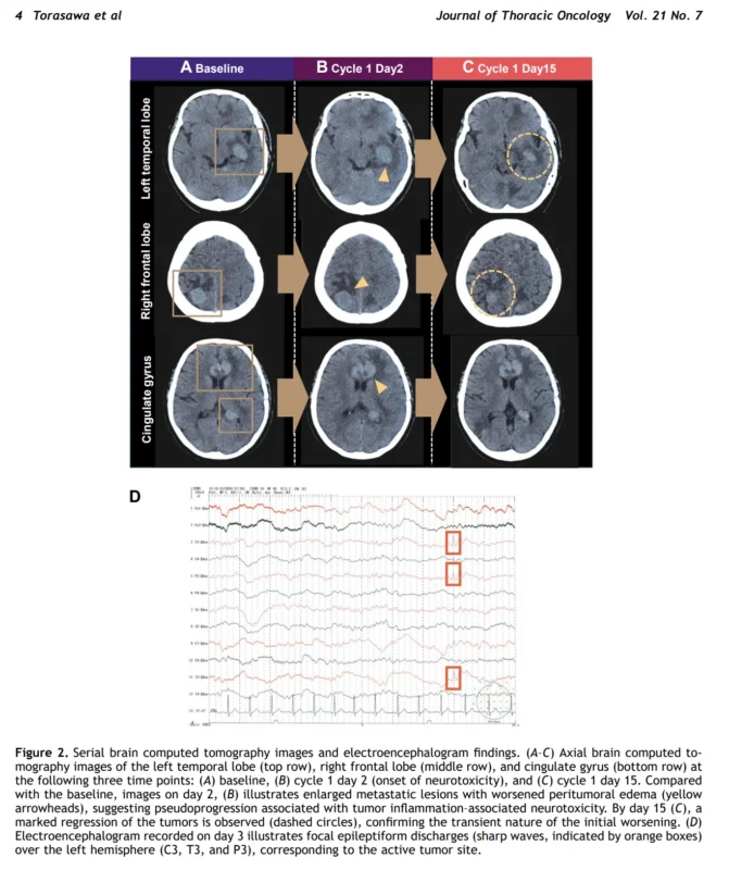
Emergency CT imaging demonstrated enlargement of existing brain metastases and worsening peritumoral edema. The initial clinical concern was severe ICANS.
She received high-dose dexamethasone, increased levetiracetam, and glycerol. Fever developed on day 3, after the neurologic event, and tocilizumab was administered.
Imaging and EEG Suggested a Localized Process
The case became more complex once the neurologic pattern was assessed in detail.
EEG performed on day 3 showed focal epileptiform discharges over the left hemisphere, corresponding to the largest active tumor site in the left temporal lobe.
This pattern differed from the generalized slowing more commonly associated with diffuse encephalopathy in ICANS.
The timing and sequence of symptoms were also relevant. The patient developed nausea and vomiting before the seizure and loss of consciousness, suggesting raised intracranial pressure or worsening focal edema. Fever did not precede the neurologic event, as is often observed in cytokine release syndrome-associated systemic immune activation.
The imaging findings also showed transient enlargement of lesions and worsening peritumoral edema. By day 15, however, CT imaging showed marked regression of multiple brain metastases.
The study’s Figure 2 illustrates this sequence clearly: early radiographic worsening at the onset of neurotoxicity was followed by regression of several intracranial lesions after treatment continuation.
This pattern supported pseudoprogression caused by local immune-mediated tumor inflammation.
What Is Tumor Inflammation-Associated Neurotoxicity?
TIAN was first described in patients with CNS lymphoma treated with CAR T-cell therapy.
Unlike ICANS, which is generally associated with diffuse cerebral dysfunction and systemic immune activation, TIAN is thought to result from localized inflammation within or around CNS tumors.
TIAN can cause worsening edema, focal deficits, seizures, increased intracranial pressure, transient mass effect, and radiographic pseudoprogression.
The clinical features reported in this patient were consistent with a TIAN-dominant process:
- Hyperacute onset within hours of treatment
- Nausea and vomiting before neurologic deterioration
- Focal-to-bilateral seizures
- Worsening local edema around brain metastases
- Focal epileptiform activity on EEG
- Rapid recovery after corticosteroids and antiseizure management
- Early reduction in tumor burden after transient radiographic worsening
Her serum progastrin-releasing peptide level also declined from 9409 pg/mL at baseline to 4878 pg/mL by day 6, providing an additional signal of antitumor activity.
Rapid Recovery Allowed a Cautious Rechallenge
The patient’s neurologic status returned to baseline within 36 hours.
After recovery, she had no aphasia, agraphia, or other persistent features that might support diffuse ICANS. There were also no new neurologic deficits attributable to the brain metastases.
Based on the rapid resolution, focal EEG findings, tumor-associated imaging changes, and early treatment response, the treating team judged the event to be more consistent with TIAN than with pure ICANS.
Tarlatamab was restarted at 1 mg on day 9 and escalated to 10 mg on day 16.
Dexamethasone and levetiracetam were continued, while lacosamide was added as a second antiseizure medication.
No recurrent severe neurologic event was reported after tarlatamab rechallenge. The patient had marked intracranial tumor regression by day 15, and spinal cord metastases also regressed.
Why the TIAN Versus ICANS Distinction Matters
The difference between these entities has direct implications for treatment continuation.
Guidelines generally recommend permanent discontinuation after grade 4 ICANS because of the potential for severe and irreversible neurologic injury.
TIAN may be different. It may represent local inflammatory activity around a responding CNS tumor and could, in selected circumstances, be managed with corticosteroids, antiseizure therapy, close neuroimaging, EEG assessment, and careful monitoring.
This does not mean that grade 4 neurotoxicity should routinely be rechallenged.
The report describes one patient. It does not establish the safety of continuing tarlatamab after severe neurotoxicity, nor does it provide a validated diagnostic framework for distinguishing TIAN from ICANS.
However, the authors suggest that several features may support a TIAN-dominant interpretation:
- Rapid and sustained neurologic recovery
- Focal seizures or focal EEG abnormalities
- Peritumoral edema or transient lesion enlargement on imaging
- Exclusion of hemorrhage, infection, and nonconvulsive status epilepticus
- Early evidence of tumor response through imaging or biomarkers
Limits of a Single Case
This report has substantial limitations.
It describes only one patient treated at one institution. The patient also had several factors that could increase neurologic risk, including multiple active brain metastases, prior whole-brain radiotherapy, pre-existing epilepsy, residual temporal lobe disease, and baseline edema.
MRI was not obtained at the point of greatest neurologic deterioration. The clinicians relied on serial CT imaging to assess worsening edema and subsequent response.
No cerebrospinal fluid studies or prospective cytokine analyses were performed. Therefore, the biological mechanisms underlying the event cannot be confirmed.
The case should not change routine tarlatamab safety management. It does, however, support prospective research into the frequency, risk factors, imaging patterns, EEG findings, and management of TIAN in patients with SCLC brain metastases.
The Bottom Line
Severe neurologic toxicity after tarlatamab in patients with SCLC and active brain metastases may not always represent classic ICANS.
In this case, focal seizures, worsening peritumoral edema, localized EEG abnormalities, rapid corticosteroid-responsive recovery, and subsequent intracranial tumor regression supported tumor inflammation-associated neurotoxicity.
TIAN remains an emerging concept in SCLC. It should be considered only through multidisciplinary assessment involving oncology, neurology, neuroradiology, and critical care teams.
For selected patients with limited therapeutic alternatives, recognizing a possible TIAN pattern may help clinicians assess whether continued tarlatamab treatment remains feasible after a severe but reversible neurologic event.
References
- Torasawa M, Shukuya T, Huang J, et al. Tumor inflammation-associated neurotoxicity masquerading as severe immune effector cell-associated neurotoxicity syndrome in a patient with SCLC treated with tarlatamab: a case report. Journal of Thoracic Oncology. 2026;21:103649.
- Ahn MJ, Cho BC, Felip E, et al. Tarlatamab for patients with previously treated small-cell lung cancer. New England Journal of Medicine. 2023;389:2063–2075.
- Mountzios G, Sun L, Cho BC, et al. Tarlatamab in small-cell lung cancer after platinum-based chemotherapy. New England Journal of Medicine. 2025;393:349–361.
- Mahdi J, Dietrich J, Straathof K, et al. Tumor inflammation-associated neurotoxicity. Nature Medicine. 2023;29:803–810.
- Kaulen LD, Martinez-Lage M, Abramson JS, et al. Clinical presentation, management, and outcome of tumor inflammation-associated neurotoxicity in CNS lymphoma treated with CD19 CAR T-cell therapy. Blood. 2025;146:1902–1913.