Spread through air spaces, or STAS, has become an increasingly important pathological feature in early-stage lung adenocarcinoma. Defined by tumor cells extending beyond the edge of the primary lesion into adjacent alveolar spaces, STAS has been associated with recurrence, particularly after limited resection, and is now incorporated as a histologic descriptor in the ninth edition TNM framework. Yet an important question has remained unresolved: does STAS simply accompany other adverse pathological features, or does it represent a biologically distinct form of early lung cancer?
A new study in the British Journal of Cancer provides one of the most detailed molecular analyses to date addressing that question. Stephanie P. L. Saw, Mengyuan Pang, and colleagues performed integrated whole-exome and transcriptomic profiling in patients with stage I lung adenocarcinoma and found that STAS-positive tumors are characterized by greater genomic instability, enrichment of TP53 alterations, whole-genome doubling, non-TRU transcriptional phenotypes, and activation of cell-cycle and DNA-replication pathways.
Importantly, STAS remained independently associated with inferior disease-free survival after adjustment for pathological stage, surgical approach, lymphovascular invasion, TP53 status, EGFR mutation status, and adjuvant treatment. The findings therefore support STAS not simply as a microscopic pattern of tumor spread, but as a potential marker of intrinsically aggressive disease biology.
STAS Was Common Even in Stage I Disease
The analysis included 378 patients with AJCC eighth-edition stage I lung adenocarcinoma, all with known STAS and EGFR status and at least two years of postoperative follow-up. STAS was present in 205 patients, or 54.2% of the cohort. Most patients underwent lobectomy, and approximately three-quarters of tumors were EGFR mutated.
The high prevalence itself is clinically relevant. STAS is not an uncommon pathological curiosity confined to a very small subgroup of aggressive tumors. In this cohort, it was present in more than half of resected stage I adenocarcinomas. Its distribution was similar according to EGFR mutation status, suggesting that STAS is not simply a manifestation of one particular oncogenic driver.
STAS-Positive Tumors Had Multiple Adverse Pathological Features
STAS-positive tumors were significantly enriched for several established markers of aggressive disease.
Stage IB disease was present in 40.0% of STAS-positive tumors compared with 22.0% of STAS-negative tumors. Lymphovascular invasion was observed in 30.7% versus 9.2%, while poorly differentiated histology was seen in 18.5% versus 2.9%. Micropapillary or solid-predominant adenocarcinoma was also much more frequent, at 10.7% versus 1.2%.
PD-L1 expression was also higher: 46.4% of STAS-positive tumors had PD-L1 TPS ≥1% compared with 27.2% of STAS-negative tumors. These associations could have suggested that STAS was simply acting as a surrogate for known high-risk pathology. The survival analysis showed otherwise.
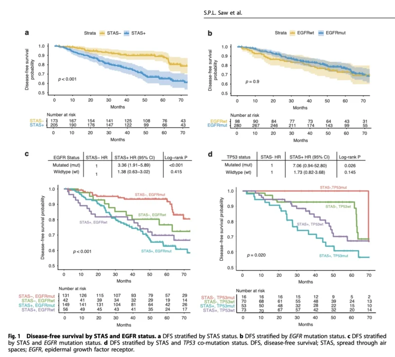
Five-Year DFS Was 65% With STAS Versus 89% Without STAS
Patients with STAS-positive tumors experienced substantially poorer disease-free survival. Estimated 2-year DFS was:
- 85.5% with STAS
versus
- 94.0% without STAS.
By five years, the difference had widened considerably:
- 65.0% versus 88.9%.
The relationship was observed within both stage IA and stage IB disease, suggesting that STAS provides information beyond conventional T-stage subdivision. This is clinically important because stage I lung cancer is often discussed as a relatively favorable disease category, yet the data show that biological heterogeneity within stage I remains substantial.
A stage IA tumor with STAS may therefore not behave like another stage IA tumor without it.
STAS Remained Independently Prognostic
The strongest clinical evidence came from multivariable analysis. After adjustment for stage, surgical approach, TP53 mutation, lymphovascular invasion, EGFR mutation status and adjuvant therapy, STAS remained independently associated with inferior DFS:
- HR 2.32; 95% CI, 1.16–4.63; P=0.017.
Sublobar resection was also independently associated with worse DFS, with an HR of 2.64. This finding strengthens the argument that STAS is not merely correlated with aggressive pathology. It appears to contribute prognostic information even after those factors are considered. The observation is especially relevant to surgical decision-making, because previous studies have consistently suggested that limited resection may be less suitable when STAS is present.
EGFR Mutation Did Not Eliminate the Prognostic Effect
One of the most interesting aspects of the study was the analysis according to EGFR status. Overall DFS did not differ significantly between EGFR-mutant and EGFR-wildtype tumors. However, among patients with EGFR-mutant stage I lung adenocarcinoma, STAS was associated with a particularly pronounced survival difference.
Five-year DFS was:
- 62.8% in STAS-positive EGFR-mutant disease
versus
- 93.4% in STAS-negative EGFR-mutant disease.
A similar direction of effect was seen in EGFR-wildtype tumors, although statistical significance was not reached, probably because that subgroup was considerably smaller. These findings suggest that STAS may identify biologically aggressive disease within an oncogene-defined population that might otherwise be regarded as relatively homogeneous.
That has potential implications for the evolving use of perioperative EGFR-targeted therapy.
TP53 Co-Mutation Was Strongly Enriched in STAS
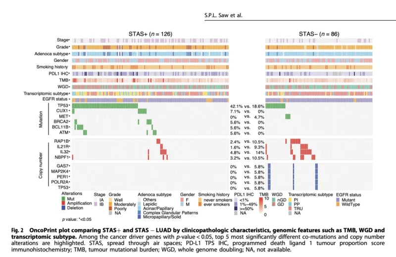
Integrated molecular profiling was available for 212 patients. One of the clearest genomic findings was enrichment of TP53 mutations in STAS-positive tumors:
- 42.1% with STAS versus 18.6% without STAS.
TP53 alterations are already recognized as markers of more aggressive biology in several oncogene-driven NSCLC subsets. The relationship between STAS and TP53 was therefore particularly interesting. The adverse effect of STAS on DFS appeared more consistently among TP53-mutated tumors, although subgroup numbers were relatively small and the authors appropriately caution that this interaction requires further validation.
Importantly, however, STAS remained independently prognostic even after TP53 status was included in the multivariable model. So TP53 enrichment does not fully explain the adverse phenotype.
Whole-Genome Doubling Points to Greater Genomic Instability
STAS-positive tumors also demonstrated greater overall genomic instability. Whole-genome doubling was observed in:
- 59.5% of STAS-positive tumors
versus
- 41.9% of STAS-negative tumors.
Tumor mutational burden was also modestly but significantly higher.
Additional alterations enriched in STAS included CUX1, BRCA2, BCL11B and ATM, although these occurred at relatively low frequencies and should not currently be interpreted as individual therapeutic biomarkers. The broader pattern is more informative than any single alteration.
STAS-positive tumors appear genomically less stable and more evolutionarily complex. The enrichment of DNA-damage repair gene abnormalities together with whole-genome doubling provides a molecular framework that may help explain their more aggressive clinical behavior.
Transcriptomics Reveals a Shift Away From the TRU Phenotype
The transcriptomic analysis added another layer to the biology. Lung adenocarcinoma can be categorized into transcriptional phenotypes including terminal respiratory unit, proximal proliferative and proximal inflammatory subtypes.
STAS-positive tumors demonstrated a marked shift toward the more aggressive non-TRU phenotypes. Proximal proliferative or proximal inflammatory transcriptional subtypes were present in 55.7% of STAS-positive tumors compared with 23.3% of STAS-negative tumors.
Conversely, the TRU phenotype accounted for 76.7% of STAS-negative tumors but only 44.3% of STAS-positive tumors. This finding supports the idea that STAS reflects an underlying biological state rather than simply a pattern produced during specimen handling or a passive consequence of tumor architecture.
Cell-Cycle and DNA-Replication Programs Were Activated
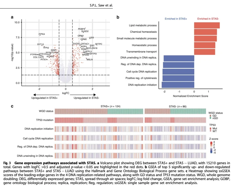
Gene-expression analysis provided perhaps the clearest mechanistic signal. STAS-positive tumors were enriched for pathways involved in: DNA replication, cell-cycle progression, DNA unwinding, replication initiation and cytokinesis. STAS-negative tumors, in contrast, showed greater enrichment of metabolic and homeostatic pathways.
The figure on page 6 of the paper illustrates this separation particularly well: DNA-replication and cell-cycle pathways cluster on the STAS-positive side, while lipid metabolism, chemical homeostasis and other metabolic processes are enriched among STAS-negative tumors.
Importantly, activation of cell-cycle pathways remained evident after stratification by TP53 status. This suggests that the transcriptomic phenotype cannot be explained solely by the greater prevalence of TP53 mutations.
STAS May Represent a Histologic Manifestation of Aggressive Tumor Biology
Taken together, the genomic and transcriptomic findings support a coherent biological model.
STAS-positive tumors are more likely to demonstrate high-grade morphology and lymphovascular invasion; at the molecular level, they show greater genomic instability, TP53 alterations, whole-genome doubling and activation of proliferative pathways.
The authors therefore propose that STAS may be a histologic manifestation of an underlying aggressive molecular state, rather than simply reflecting how tumor cells physically disperse into adjacent alveoli. This is probably the most important conceptual contribution of the study.
Histology and molecular oncology are often treated as separate domains. STAS may represent a feature where the two converge: a microscopic pattern that visually identifies a biologically aggressive tumor phenotype.
The Findings Could Influence Surgical Risk Assessment
STAS already has implications for surgery. Multiple retrospective series have shown higher recurrence after sublobar resection when STAS is present, and the present study again found sublobar resection independently associated with inferior DFS.
The challenge is that STAS is usually diagnosed on the resection specimen, when the extent of surgery has already been decided. That limits its immediate ability to guide the original surgical approach.
Future research may therefore need to determine whether preoperative imaging, frozen-section pathology, radiomics or other biomarkers can reliably predict STAS before or during surgery. If that becomes possible, STAS status could contribute to the choice between segmentectomy, wedge resection and lobectomy in carefully selected stage I disease.
Could STAS Help Select Patients for Adjuvant Therapy?
The more provocative implication relates to postoperative systemic therapy.
Most stage IA tumors and many stage IB tumors are treated primarily with surgery. Yet the markedly poorer DFS observed among STAS-positive patients raises the possibility that STAS could identify a biologically high-risk subgroup within conventional early-stage categories.
The authors specifically highlight the need to investigate whether STAS-positive stage I disease should influence adjuvant treatment selection. The enrichment of DNA-damage repair alterations and proliferative signaling also raises the hypothesis that some STAS-positive tumors might be more sensitive to platinum-based chemotherapy.
But this remains entirely prospective. The study does not demonstrate that chemotherapy improves outcomes specifically in STAS-positive stage I disease, and adjuvant treatment should not currently be prescribed solely because STAS is present.
The EGFR-Mutant Population Creates Another Important Question
The finding may have particular relevance in EGFR-mutant disease. Adjuvant osimertinib is already established after resection in eligible EGFR-mutated NSCLC, while current research is exploring its role in progressively earlier stages.
If STAS identifies an EGFR-mutant stage I subgroup with markedly higher recurrence risk, histologic features such as STAS may eventually complement pathological stage and molecular biomarkers when determining who requires postoperative targeted therapy.
This idea aligns closely with the broader movement toward risk-adapted adjuvant treatment in EGFR-mutant NSCLC. Rather than treating all tumors within a stage as biologically equivalent, future strategies may combine anatomical stage, STAS, genomic co-mutations, transcriptomic risk, and postoperative molecular residual disease.
Important Limitations Prevent Immediate Clinical Translation
The study is retrospective and originates from a single Singapore institution. Approximately 74% of tumors were EGFR mutated, substantially higher than would be expected in many Western lung cancer populations. The relative underrepresentation of EGFR-wildtype tumors and other molecular subsets—including KRAS G12C and ALK-positive disease, limited subgroup analyses.
Only 212 patients underwent comprehensive WES and RNA sequencing. The number of recurrence events was also modest, and detailed analyses of recurrence patterns were limited. External validation of the molecular findings was constrained by the scarcity of stage I datasets combining annotated STAS status with genomic and transcriptomic profiling.
Most importantly, the study demonstrates association rather than causation. It cannot determine whether the molecular abnormalities produce STAS, whether STAS facilitates aggressive behavior, or whether both arise from a common underlying biological program. Spatial transcriptomics and prospectively collected multicenter cohorts may help answer that question.
The Bottom Line
This British Journal of Cancer study provides strong evidence that STAS is more than a histologic descriptor in stage I lung adenocarcinoma. Among 378 patients, STAS was present in 54.2% and was associated with markedly worse DFS. Five-year DFS was:
- 65.0% with STAS
versus
- 88.9% without STAS.
After multivariable adjustment, STAS remained independently associated with recurrence or death:
- HR 2.32.
The molecular analysis provides a plausible biological explanation. STAS-positive tumors were enriched for TP53 mutations, whole-genome doubling, non-TRU transcriptional phenotypes, higher PD-L1 expression and activation of DNA-replication and cell-cycle programs.
The clinical implication is not that STAS should immediately dictate adjuvant treatment. It is that stage I lung adenocarcinoma cannot be considered biologically uniform.
As early-stage lung cancer becomes increasingly molecularly stratified, STAS may ultimately function as a bridge between pathology and precision oncology—helping identify tumors whose aggressive biology is not fully captured by stage alone.
Reference
- Saw SPL, Pang M, Patteri C, Takano A, Yeo JC, Sim NL, Lai GGY, Lim DWT, Ang M-K, Kanesvaran R, Ng QS, Jain A, Tan WL, Tan AC, Tan WC, Seet AOL, Ong B-H, Lim TKH, Skanderup AJ, Tan DSW. Genomic and transcriptomic landscape of spread through air spaces in stage 1 lung adenocarcinoma. British Journal of Cancer. 2026. doi:10.1038/s41416-026-03622-8.