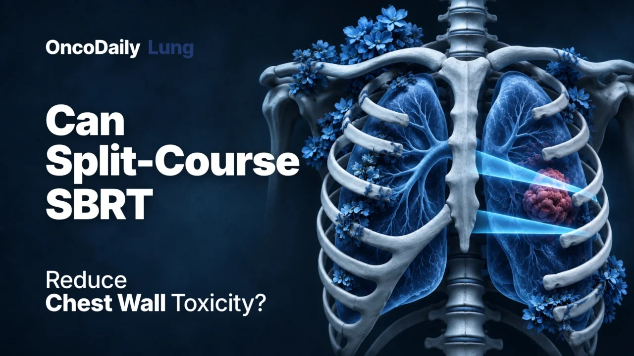
A 2026 case report published in Medical Dosimetry describes an innovative stereotactic body radiotherapy (SBRT) planning strategy designed to reduce radiation exposure to the ribs and chest wall when treating pulmonary lesions located close to critical structures. By dividing treatment between inspiration and expiration breath-hold positions, investigators demonstrated the feasibility of reducing dose to adjacent healthy tissues while delivering an ablative radiotherapy course. Although promising, the approach remains preliminary and requires further clinical validation.
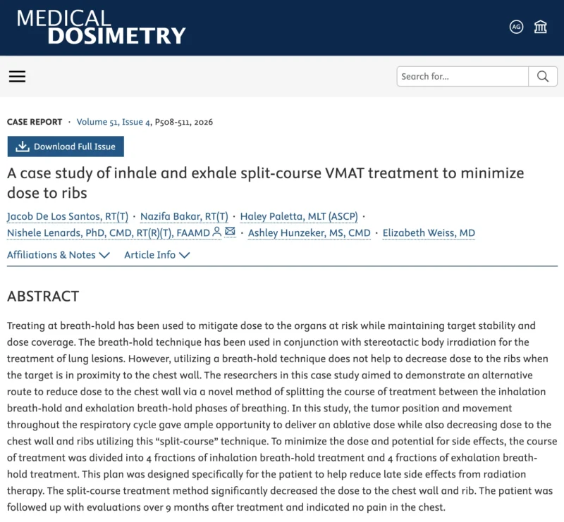
The study, titled A Case Study of Inhale and Exhale Split-Course VMAT Treatment to Minimize Dose to Ribs, was conducted by Jacob De Los Santos, Nazifa Bakar, Haley Paletta, Nishele Lenards, Ashley Hunzeker, and Elizabeth Weiss.
Published in Medical Dosimetry in 2026, the report describes an individualized volumetric modulated arc therapy (VMAT) approach for an 84-year-old woman with renal cell carcinoma and a 2.1-cm pulmonary metastasis adjacent to the chest wall.
The central innovation was to distribute treatment across two respiratory positions rather than use one breath-hold position throughout the entire course. Four fractions were delivered during inspiration breath-hold and four during expiration breath-hold.
The investigators reported reduced radiation dose to the adjacent ribs and chest wall, and the patient experienced no chest pain during nine months of follow-up.
These findings introduce an interesting strategy for managing organ-at-risk exposure in thoracic stereotactic radiotherapy. However, they do not establish clinical superiority or demonstrate that rib fractures can be prevented.
The Challenge of Chest Wall Toxicity in Lung SBRT
SBRT has become an essential component of modern thoracic radiation oncology. Its ability to deliver highly conformal ablative radiation doses over a limited number of fractions makes it particularly valuable in patients with early-stage lung cancer who are medically unsuitable for surgery.
The technique also has an established role in selected patients with pulmonary metastatic disease. Unlike conventional radiotherapy, SBRT delivers high biological doses while achieving a steep dose gradient outside the target volume.
However, the proximity of pulmonary lesions to surrounding anatomical structures can limit treatment planning. Relevant organs at risk include normal lung tissue, the esophagus, heart, spinal cord, chest wall, and ribs.
For tumors located directly adjacent to the chest wall, the challenge becomes particularly important because the target and surrounding bony structures may receive overlapping high-dose distributions. Radiation-associated rib fractures and chest wall pain represent recognized late complications of thoracic SBRT.
Such complications may produce chronic pain, reduced mobility, and impaired quality of life. The risk depends on several factors, including tumor-to-chest-wall distance, radiation dose distribution, treated volume, and patient characteristics.
In a pooled analysis of 57 studies, the estimated incidence of rib fracture after thoracic SBRT was approximately 6.3%, while chest wall pain occurred in approximately 11.0%. However, reported fracture rates vary considerably across treatment settings and studies.
Patients whose tumors directly abut the chest wall may face greater risks than those with more centrally located lesions. These observations highlight the need for techniques that preserve tumor dose while limiting exposure to vulnerable adjacent tissues.
Why Conventional Breath-Hold Techniques May Not Be Sufficient
Respiratory motion presents a major challenge during lung radiotherapy. With normal breathing, pulmonary tumors move relative to surrounding anatomy. This movement can complicate target localization and increase the volume of tissue receiving radiation.
Breath-hold techniques are commonly used to improve target reproducibility and reduce respiratory motion during treatment delivery. Deep inspiration breath-hold can increase lung volume and change the spatial relationship between the target and surrounding structures.
However, breath-hold treatment does not automatically reduce rib or chest wall dose when a tumor lies immediately adjacent to these tissues. In some patients, even a highly conformal plan may expose neighboring ribs to substantial radiation because the tumor and chest wall remain closely positioned.
The researchers therefore investigated whether the changing anatomical relationship between the tumor and ribs during inspiration and expiration could be used more effectively. Rather than selecting a single respiratory position for all fractions, they proposed distributing treatment across two breath-hold positions.
A New Strategy: Splitting Treatment Between Inspiration and Expiration
The central concept of the study was the use of separate treatment plans corresponding to inspiration and expiration breath-hold. Under conventional breath-hold SBRT, all treatment fractions are generally delivered using a selected respiratory position.
In the proposed split-course technique, the full course was divided between two respiratory configurations. Four fractions were delivered during inspiration breath-hold and four during expiration breath-hold.
Because the tumor and chest wall change their relative positions during the breathing cycle, the investigators aimed to alter how high-dose regions were distributed across adjacent structures. In principle, this approach may reduce the concentration of cumulative radiation dose within the same rib segments.
The strategy requires careful planning to ensure that the prescribed tumor dose is maintained while the cumulative dose to normal tissues remains within acceptable limits. Crucially, this approach should not be confused with a split-course treatment interrupted by a prolonged interval.
Here, the term describes dividing radiotherapy delivery between two respiratory positions. The technique represents a form of individualized motion management and dose distribution rather than a change to the fundamental radiobiological objectives of SBRT.
The Clinical Case: A Pulmonary Metastasis Adjacent to the Chest Wall
The report describes an 84-year-old woman with a history of renal cell carcinoma who developed a 2.1-cm metastatic lesion in the right lower lung. The lesion was located close to the chest wall, making dose exposure to adjacent ribs an important treatment-planning consideration.
The patient was receiving oncological management that included systemic therapy and radiotherapy. The treating radiation oncology team selected a breath-hold-based stereotactic approach.
The investigators compared the potential radiation exposure associated with a conventional single-position breath-hold plan against a strategy using both inspiration and expiration breath-hold configurations.
The aim was to preserve appropriate ablative treatment delivery while reducing dose to the chest wall and adjacent ribs.
The choice of the two breathing positions was particularly relevant because the patient’s tumor location and respiratory motion provided an opportunity to modify the relative geometry between the target and nearby bony structures. This anatomic relationship was central to the reported planning benefit.
Dosimetric Findings: Reduced Exposure to Ribs and Chest Wall
The authors reported that the split-course VMAT technique decreased radiation dose to the chest wall and ribs compared with the conventional breath-hold planning approach. This finding supports the underlying hypothesis that respiratory-position changes may be exploited to redistribute radiation exposure.
However, the publicly available abstract does not provide the precise maximum rib dose, absolute dose reduction in Gy, or a complete comparison of cumulative dose-volume parameters. Consequently, the magnitude of the dosimetric improvement cannot be independently quantified from the abstract alone.
The report also does not provide evidence that the technique improves local tumor control compared with standard SBRT. The central demonstrated outcome is the feasibility of an individualized planning approach that the investigators found capable of reducing exposure to selected organs at risk.
The distinction between dosimetric improvement and clinical benefit is essential. Although reducing high-dose exposure to ribs is biologically and clinically reasonable, a favorable treatment plan does not necessarily translate into a measurable reduction in late rib fractures or chronic chest wall pain.
Nine-Month Follow-Up: No Chest Wall Pain Reported
The patient underwent follow-up assessments over nine months after treatment. During this period, no chest pain was reported. This is an encouraging clinical observation. However, the follow-up period is relatively short for assessing radiation-associated rib fractures. Late rib toxicity may appear many months or even years after stereotactic treatment.
A previous radiological study of thoracic stereotactic radiotherapy reported that rib fractures appeared at a mean of approximately 21 months after treatment. Therefore, the absence of pain at nine months cannot establish long-term safety.
Furthermore, rib fractures may be asymptomatic and detected only during imaging surveillance. The case report does not demonstrate that the new technique prevents rib fractures, reduces long-term toxicity rates, or provides superior quality of life.
These questions require longer follow-up and evaluation in larger patient populations.
Clinical Implications: Could Respiratory Motion Become a Planning Advantage?
The proposed strategy introduces an interesting change in how respiratory motion may be considered during thoracic radiotherapy planning. Traditionally, respiratory motion is primarily treated as a source of uncertainty requiring control or compensation.
Breath-hold, gating, tracking, and motion-encompassing approaches seek to ensure accurate target coverage while reducing unintended exposure to surrounding tissues. The split-course strategy suggests that respiratory variation may also provide an opportunity for dosimetric optimization.
In selected patients, inspiration and expiration can create different spatial relationships between a target and nearby organs at risk. If those differences are sufficiently reproducible, treatment could potentially be distributed across respiratory positions to reduce cumulative dose concentration in specific tissues.
However, implementation would require accurate image registration, reproducible breath-hold performance, reliable target localization, and rigorous evaluation of the cumulative dose distribution.
The technique may be particularly relevant for selected peripheral pulmonary lesions abutting the chest wall. Its potential applicability to primary lung cancer remains a research question, because the reported patient had a pulmonary metastasis from renal cell carcinoma rather than primary NSCLC.
Limitations and Future Research
The most important limitation of the study is its single-patient design. The reported dosimetric benefit depended on the patient’s individual anatomy and tumor movement. Not all pulmonary lesions exhibit sufficient displacement relative to adjacent ribs to create a meaningful planning advantage.
Additionally, a technique requiring two respiratory configurations may introduce operational complexity, including additional simulation, planning, image guidance, and quality assurance. The reproducibility of both breath-hold positions would need to be demonstrated throughout treatment.
Dose accumulation across different anatomical positions also requires careful consideration. Further studies should quantify the maximum dose to individual ribs, relevant chest wall dose-volume parameters, normal-lung exposure, and target coverage compared with conventional approaches.
Longer-term clinical assessment should include rib fracture incidence, chest wall pain, pulmonary toxicity, and local control. A prospective planning study involving a larger group of patients would be a logical next step before evaluating clinical toxicity outcomes.
OncoDaily Takeaway
This 2026 case report introduces a potentially useful approach to reducing rib and chest wall radiation exposure during stereotactic treatment of pulmonary lesions close to the chest wall.
By dividing eight treatment fractions between inspiration and expiration breath-hold positions, investigators reported improved dose sparing in an 84-year-old patient with a renal cell carcinoma lung metastasis. The patient experienced no chest pain during nine months of follow-up.
However, this remains a feasibility and dosimetric observation from a single case. The technique has not demonstrated superiority over conventional SBRT, and its ability to reduce late rib fractures has not been established.
The central clinical implication is that respiratory motion may offer opportunities for individualized dose optimization, but rigorous dosimetric validation and longer-term clinical studies are required before this approach can become an established treatment strategy.
Sources
- De Los Santos J, Bakar N, Paletta H, Lenards N, Hunzeker A, Weiss E. A Case Study of Inhale and Exhale Split-Course VMAT Treatment to Minimize Dose to Ribs. Medical Dosimetry. 2026;51(4):508–511. DOI: 10.1016/j.meddos.2026.05.008
- Bongers EM, Haasbeek CJA, Lagerwaard FJ, Slotman BJ, Senan S. Incidence and Risk Factors for Chest Wall Toxicity After Risk-Adapted Stereotactic Radiotherapy for Early-Stage Lung Cancer. Journal of Thoracic Oncology. 2011;6:2052–2057. DOI: 10.1097/JTO.0b013e3182307e74
- Chest Wall Toxicity After Stereotactic Body Radiation Therapy: A Pooled Analysis of 57 Studies. International Journal of Radiation Oncology, Biology, Physics. 2019.
- Rib Fracture After Stereotactic Radiotherapy on Follow-Up Thin-Section Computed Tomography in 177 Primary Lung Cancer Patients. 2011.