
Pulmonary nodules are among the most common, and potentially consequential, findings encountered on chest computed tomography. Their increasing detection reflects both the widespread use of CT imaging and the expansion of lung cancer screening, but the central clinical challenge has remained unchanged: identifying the relatively small proportion of nodules that represent lung cancer without subjecting large numbers of patients with benign disease to unnecessary imaging, invasive procedures, anxiety, and cost.
A new New England Journal of Medicine Clinical Practice review by Matthew E.J. Callister and Gerard A. Silvestri provides a contemporary framework for navigating this problem. Published on September 3, 2026, the article emphasizes that pulmonary nodule management cannot be reduced to a single size threshold. Morphology, growth, comparison with prior imaging, estimated probability of malignancy, patient fitness, and preferences must be integrated before deciding whether surveillance, PET-CT, biopsy, or definitive treatment is appropriate.
The underlying principle is straightforward but clinically important: the objective is not to biopsy every nodule or to observe every indeterminate lesion. It is to identify when the probability of cancer has become high enough that additional surveillance creates more risk than benefit.
Morphology Is the Starting Point
Pulmonary nodules are conventionally divided into solid and subsolid lesions, with subsolid nodules further classified as pure ground-glass or part-solid. This distinction is fundamental because the natural history and management of these lesions differ substantially.
Solid nodules obscure the underlying bronchovascular structures, whereas pure ground-glass nodules retain visible lung architecture through the area of increased attenuation. Part-solid nodules contain both ground-glass and solid components and, when malignant, may represent different stages along the spectrum from preinvasive adenocarcinoma to invasive disease.
Subsolid nodules often grow more slowly than solid nodules, yet persistent part-solid lesions can carry a particularly high probability of malignancy. Consequently, slower growth should not be confused with lower oncologic significance. The review emphasizes that the development or progressive enlargement of a solid component within a previously ground-glass lesion is one of the most important radiologic changes suggesting invasive transformation.
This distinction also explains why the duration of surveillance differs. A solid nodule that remains stable for two years can generally be considered benign, whereas subsolid nodules require longer observation, often at least four years, because malignant lesions may demonstrate much slower growth.
Copyright © 2026 Massachusetts Medical Society.
Previous Imaging May Be the Most Valuable Diagnostic Test
Before calculating malignancy risk or requesting PET-CT, the first step should be comparison with prior imaging whenever available.
Stability, growth rate, and morphological evolution can substantially change the estimated probability of malignancy. For solid nodules, two years of stability is generally reassuring enough to end surveillance, and volumetric CT may permit discharge after one year when true volumetric stability has been established.
Conversely, growth during follow-up shifts the clinical interpretation. The review notes that an increase of approximately 1.5–2 mm in diameter or about 25% in volume can be used as evidence of growth, although volumetric assessment may improve sensitivity at the expense of some specificity.
The principle is particularly important for subsolid lesions. A stable pure ground-glass nodule may remain appropriate for prolonged surveillance, whereas the appearance of a new solid component fundamentally changes its risk profile.
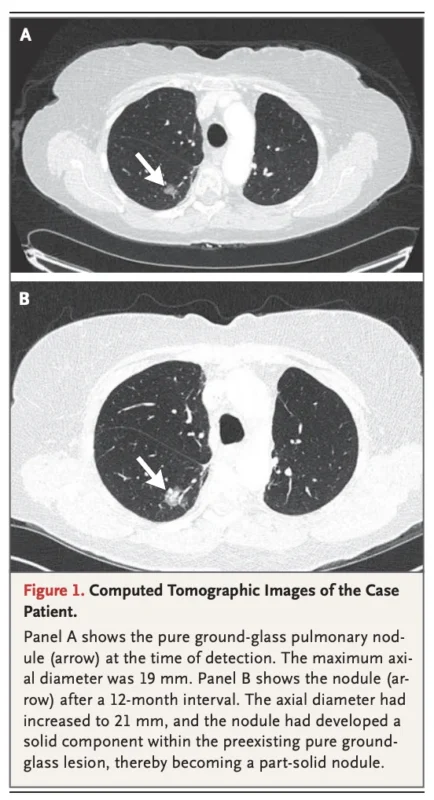
The case vignette accompanying the review illustrates this evolution particularly well. A fit 67-year-old man initially had a 19-mm pure ground-glass nodule. At six months the lesion remained stable, but at 12 months it had increased to 21 mm and developed a solid component, converting it into a part-solid nodule.
The CT images on page 3 visually demonstrate why morphology matters as much as size: the absolute increase in diameter is modest, but the emergence of a solid component represents a much more important biological change.
Solid Nodules Should Be Managed According to Malignancy Risk
For solid nodules that cannot be dismissed on the basis of benign morphology, small size, or documented stability, the next step is estimation of the probability of cancer.
Several validated tools are available, including the Mayo Clinic, Veterans Affairs, Brock, Herder, BIMC, and Cleveland Clinic models. These incorporate different combinations of age, smoking history, nodule diameter, spiculation, location, emphysema, previous cancer history, FDG avidity, and other clinical or radiologic variables.
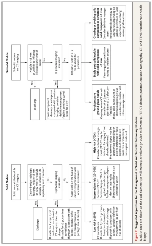
The review organizes management into clinically useful risk groups rather than relying on a single universal algorithm.
For nodules with a low estimated malignancy risk below approximately 10%, CT surveillance is generally appropriate. These lesions are often too small for reliable PET characterization or nonsurgical biopsy.
For an intermediate probability of approximately 10%–70%, PET-CT and/or tissue sampling can meaningfully refine the diagnosis.
For nodules with a high probability of malignancy above approximately 70%, definitive management may be more appropriate, and selected patients can proceed to surgical treatment without requiring preoperative biopsy.
This framework is useful because it links diagnostic intensity to pretest probability. A test has very different value in a patient with a 5% probability of cancer than in one whose probability is already above 90%.
PET-CT Is Most Useful in the Intermediate-Risk Setting
PET-CT remains an important tool for indeterminate solid nodules large enough to be meaningfully characterized.
In a meta-analysis involving more than 8,500 nodules cited by the authors, PET-CT demonstrated a sensitivity of approximately 89% and specificity of 75%. When the pretest prevalence of malignancy is 10%, a positive PET result raises the post-test probability to approximately 28%, whereas a negative result reduces it to about 1.6%.
This illustrates why PET-CT is most useful when the pretest probability is neither extremely low nor already very high. It can move an intermediate-risk lesion sufficiently in either direction to influence the decision between surveillance, biopsy, and definitive treatment.
The situation is different for evolving subsolid nodules. Because malignant subsolid lesions may be relatively indolent metabolically, absence of FDG uptake cannot reliably exclude malignancy. The review explicitly cautions that a negative PET-CT should not discourage definitive treatment when a part-solid nodule is developing or its solid component is enlarging.
PET-CT may still be useful in such cases for staging, rather than for determining whether the lesion itself is malignant.
Subsolid Nodules Require Patience, Until Their Biology Changes
The management of subsolid nodules requires a different balance between surveillance and intervention.
Some newly detected subsolid lesions are transient inflammatory or infectious abnormalities. This explains why repeat CT after approximately three to six months is commonly recommended to confirm persistence before committing a patient to prolonged surveillance or invasive investigation.
Persistent pure ground-glass nodules frequently follow an indolent course and can often remain under surveillance. Long-term follow-up is necessary because growth can occur slowly, and the authors recommend at least four years of stability before considering a subsolid lesion benign.
The threshold for intervention changes when a solid component emerges.
A previously pure ground-glass lesion that develops a solid component, or a part-solid lesion whose solid component progressively enlarges, should be regarded as being at substantial risk for invasive adenocarcinoma. In a surgically fit patient, definitive management should therefore be considered rather than continuing routine surveillance.
Persistent part-solid nodules with a solid component of approximately 8 mm or greater also warrant further investigation even if obvious interval growth has not occurred.
The management algorithm on page 9 captures this distinction clearly: stable pure ground-glass lesions remain candidates for prolonged CT surveillance, whereas an evolving solid component shifts the pathway toward biopsy or definitive treatment.
The Biopsy Decision Is Becoming More Nuanced
When tissue diagnosis is appropriate, transthoracic needle biopsy and navigational bronchoscopy remain the principal nonsurgical approaches. The decision depends on nodule size and location, local expertise, pneumothorax risk, and patient preference.
Historically, transthoracic biopsy was generally assumed to have substantially higher diagnostic accuracy. Older meta-analyses reported an accuracy of approximately 92%, although pneumothorax occurred in about 20% of procedures. Guided bronchoscopy historically produced lower diagnostic yields but substantially fewer pneumothoraces.
More recent randomized evidence has narrowed that gap.
In the trial discussed in the NEJM review, diagnostic accuracy was 74% with transthoracic needle biopsy and 79% with navigational bronchoscopy. The difference in complications was much more pronounced: pneumothorax occurred in 28% versus 3%, respectively, while chest-tube placement, hospitalization, or both occurred in 12% versus 1%.
These findings make navigational bronchoscopy increasingly attractive when both techniques are technically feasible.
Robotic-assisted bronchoscopy may further expand access to peripheral lesions, although its incremental diagnostic benefit remains uncertain. Published studies have produced variable yields, and the NEJM authors appropriately avoid presenting robotic bronchoscopy as uniformly superior to established guided bronchoscopic approaches.
A Negative Biopsy Does Not Necessarily Mean a Benign Nodule
The value of biopsy must always be interpreted in relation to pretest probability. This becomes particularly important in lesions that already have a very high probability of malignancy.
The authors provide a useful example: if the pretest probability of malignancy is 95%, a nonspecific or nondiagnostic robotic bronchoscopic biopsy may still leave an estimated 80% probability of cancer.
In this situation, a nondiagnostic biopsy does not meaningfully resolve the clinical question and may simply delay definitive management.
This explains why preoperative biopsy is not mandatory for every high-risk nodule. In a medically fit patient with a highly suspicious, surgically resectable lesion, proceeding directly to diagnostic and therapeutic resection may be appropriate after multidisciplinary discussion and shared decision-making.
Conversely, biopsy becomes especially useful when a plausible benign alternative exists, when surgical risk is substantial, or when the planned treatment is nonsurgical and histologic confirmation is required.
Surgery Is Also Evolving Toward Lung Preservation
For decades, lobectomy represented the standard surgical treatment for stage I lung cancer. Contemporary evidence has expanded the role of sublobar resection for appropriately selected small peripheral tumors.
The review notes randomized evidence demonstrating similar disease-free and overall survival with sublobar resection and lobectomy for lesions measuring 2 cm or less. Lung preservation can be particularly relevant for patients with limited cardiopulmonary reserve, synchronous pulmonary tumors, or a future risk of developing additional primary lung cancers.
For medically inoperable patients, or those who decline surgery, stereotactic body radiotherapy provides an alternative capable of achieving good local tumor control.
The broader implication is that nodule management no longer ends simply with the question of whether to operate. Once malignancy is sufficiently likely or confirmed, clinicians must also determine the most appropriate extent and modality of definitive local treatment.
Copyright © 2026 Massachusetts Medical Society.
AI and Blood-Based Biomarkers May Refine the Indeterminate Middle Ground
The greatest unmet need remains the intermediate-risk nodule.
Patients with clearly benign lesions can be observed or discharged, while highly suspicious lesions can proceed toward definitive treatment. The difficult group lies between these extremes, where current management often requires PET-CT, biopsy, repeated surveillance, or some combination of these approaches.
Blood-based biomarkers may eventually reduce uncertainty by identifying patients who can safely remain under surveillance or those who should move more quickly toward invasive diagnosis. However, the authors emphasize that relatively few studies have yet demonstrated genuine clinical utility rather than simply diagnostic discrimination.
Artificial intelligence represents another rapidly developing approach. Machine-learning systems can integrate CT characteristics with clinical variables and potentially molecular biomarkers to estimate malignancy risk. Their promise is substantial, particularly for high-volume screening and incidental-nodule workflows, but current limitations include retrospective development, inadequate external validation, bias, lack of transparency, and difficulties integrating black-box predictions into clinical decisions.
The review therefore positions AI appropriately: as an adjunct to clinical judgment rather than its replacement.
Surveillance Also Has Risks
The harm of overdiagnosis and unnecessary invasive testing is well recognized, but surveillance is not inherently risk-free.
A malignant lesion observed for too long can undergo a stage shift, potentially moving from localized curable disease to nodal or metastatic involvement. Evidence quantifying this risk remains incomplete, yet the possibility becomes increasingly relevant as bronchoscopic technologies make smaller nodules accessible for tissue diagnosis.
The challenge is that lowering the threshold for biopsy has its own consequences. Only approximately 3%–4% of nodules identified in screening and many incidental cohorts ultimately represent cancer. More aggressive biopsy of small lesions would therefore expose substantially more patients with benign disease to procedural risk.
Optimal nodule care sits precisely between these two errors: delaying diagnosis of clinically relevant cancer and overinvestigating biologically insignificant or benign disease.
Communication Is Part of Nodule Management
The uncertainty surrounding pulmonary nodules creates substantial psychological burden. The review reports distress in approximately 40%–50% of patients undergoing evaluation, with better clinician communication associated with less distress.
Patients frequently overestimate the probability that a pulmonary nodule represents cancer, particularly when the estimated risk is never explicitly discussed.
Shared decision-making therefore becomes especially important when several guideline-concordant options exist. A patient choosing between surveillance, biopsy, and surgery should understand not simply the technical differences among those approaches, but the estimated probability of cancer, procedural risks, consequences of delayed diagnosis, and likelihood that the lesion would become clinically relevant during their lifetime.
This is particularly important for older patients with major comorbidities or limited life expectancy, in whom aggressive investigation of an indolent nodule may provide little meaningful benefit.
The Case Vignette Illustrates the Decision Point
The NEJM case brings the principles together. A 19-mm pure ground-glass nodule initially justified surveillance. Twelve months later, however, the lesion measured 21 mm and had developed a solid component.
The authors estimate the probability of invasive adenocarcinoma in this setting at greater than 95% and recommend discussing either percutaneous biopsy or diagnostic resection. If biopsy confirms lung cancer, or if the patient elects surgery without preoperative tissue confirmation, they recommend PET-CT for staging followed by robotic-assisted segmentectomy.
The important lesson is not the two-millimeter increase in diameter alone.
It is the morphological evolution from pure ground-glass to part-solid disease.
That change transforms the risk assessment and shifts management from observation toward definitive diagnosis and treatment.
The Bottom Line
Contemporary pulmonary nodule management is best understood as risk-adapted decision-making rather than size-based decision-making. Solid and subsolid nodules have different natural histories, and previous imaging remains essential for evaluating growth and morphological evolution.
Low-risk solid nodules can generally undergo CT surveillance, intermediate-risk lesions are candidates for PET-CT and/or biopsy, and highly suspicious nodules may proceed directly toward definitive management in appropriately selected patients.
Subsolid nodules require particular attention because their indolent growth can be misleading. Persistent pure ground-glass lesions may remain under long-term surveillance, but development or enlargement of a solid component should substantially increase concern for invasive adenocarcinoma and prompt consideration of definitive treatment.
At the same time, modern navigational bronchoscopy, lung-sparing surgery, emerging biomarkers, volumetric imaging, and artificial intelligence are expanding the tools available to clinicians. The objective, however, remains unchanged: diagnose clinically important lung cancer early enough to preserve curability while protecting patients with benign or indolent disease from unnecessary intervention.
That balance, not simply finding more nodules, is the central challenge of modern pulmonary nodule care.
Reference
- Callister MEJ, Silvestri GA. Pulmonary Nodules. N Engl J Med. 2026;395:894–905. Published September 3, 2026. doi:10.1056/NEJMcp2515063.