A single-center study involving 102 patients suggests that CT-guided lung biopsy and cryoablation can be performed during the same treatment session, potentially simplifying care for patients with highly suspicious small pulmonary nodules. Among 81 patients with pathology-confirmed non-small-cell lung cancer, the recorded three-year cumulative incidence of local tumor progression was 8.8%, while overall survival reached 90.8%. Published in the Journal of Vascular and Interventional Radiology, the findings support the feasibility of a combined approach but do not establish superiority over surgery, stereotactic radiotherapy, or separate biopsy and ablation procedures.
The increasing adoption of low-dose CT screening has expanded the detection of small pulmonary nodules, creating new opportunities for earlier diagnosis and potentially curative intervention. For many patients, particularly those with significant cardiopulmonary comorbidities, however, the diagnostic and therapeutic pathway remains challenging.
A suspicious lung nodule may require CT-guided biopsy, pathological assessment, staging, multidisciplinary evaluation, and a separate appointment for definitive local treatment. These steps are essential for appropriate cancer management, but repeated procedures may add complexity for patients with limited physiological reserve.
A new study by Sameer Rehman, Shreyas Naidu, and colleagues investigates whether two of these steps can safely be combined. Their research, Synchronous Percutaneous Biopsy and Cryoablation for Suspicious Solitary Pulmonary Nodules, was published online on October 22, 2026, in the Journal of Vascular and Interventional Radiology.
The investigators evaluated 102 consecutive patients who underwent CT-guided core needle biopsy followed by cryoablation during the same encounter.
The approach achieved complete intraprocedural technical success, provided a specific histopathological diagnosis in 87.3% of cases, and yielded encouraging local-control outcomes among patients subsequently confirmed to have NSCLC.
Nevertheless, the findings also highlight the importance of carefully selecting patients, particularly because definitive ablation was performed before final pathology became available.
Why a Single-Session Approach Matters
Surgical resection remains an established treatment for medically operable patients with early-stage NSCLC. For patients considered unsuitable for surgery, stereotactic body radiotherapy (SBRT) is a well-established treatment option.
Percutaneous thermal ablation offers another approach for selected patients, particularly when surgery is contraindicated and SBRT is unsuitable, carries substantial risk, or is declined. Cryoablation destroys tumor tissue through controlled freezing, resulting in cellular injury and tissue necrosis.
Unlike surgical resection, the procedure can be performed percutaneously using image-guided probes. However, conventional management often requires an initial biopsy followed by a separate treatment session.
Combining biopsy and cryoablation could potentially reduce the number of procedural encounters and avoid repeating some aspects of image-guided intervention. The central concern is that both steps must remain technically reliable.
Biopsy-related hemorrhage can obscure a small pulmonary target before the ablation probes are correctly positioned. Conversely, obtaining tissue after freezing may affect specimen quality.
The investigators therefore developed a procedural sequence intended to preserve target visualization while obtaining diagnostic tissue before therapeutic freezing.
Study Population: 102 Patients With Highly Suspicious Pulmonary Nodules
The retrospective study included patients treated at a single institution between August 2020 and October 2025. All patients had a solitary pulmonary nodule measuring between 1 and 2 cm. The cohort was highly selected. Patients had a smoking history exceeding 20 pack-years, evidence of emphysema, and imaging features considered strongly suspicious for malignancy.
Eligibility required a PET maximum standardized uptake value of at least 3.0, together with suspicious imaging morphology or interval growth. Features considered concerning included spiculated margins, lobulation, pleural indentation, and CT growth of at least 20% over six months.
Patients with radiographic nodal or distant metastatic disease were excluded. Clinical staging was based on CT and PET/CT, with endobronchial ultrasound performed selectively rather than universally.
All patients were considered medically inoperable because of cardiovascular or pulmonary risk, or had declined surgery. The mean age was 74.5 years, and the mean preprocedural FEV1 was only 35.5% of predicted.
Twelve patients had documented interstitial lung disease. The mean pulmonary nodule diameter was 1.52 cm. Most lesions were peripheral, although 40.2% were categorized as central according to the study’s anatomical definition.
These characteristics underline the clinical challenge: the investigators were treating small suspicious lesions in an older population with substantial pulmonary impairment.
The Technique: Placing a Cryoprobe Before Biopsy
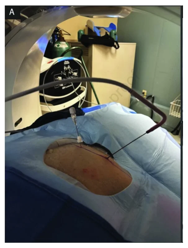
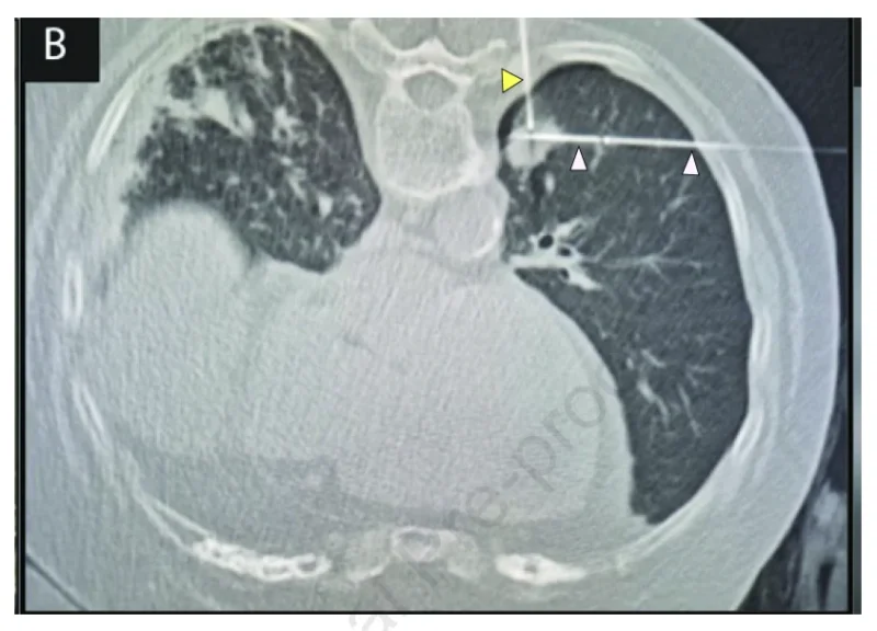
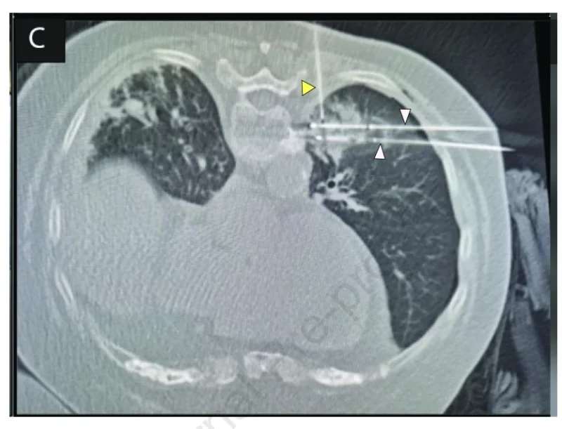
The procedural innovation was the sequence of probe placement and tissue sampling. All interventions were performed under general anesthesia with CT guidance by an experienced thoracic interventional radiologist. First, a cryoprobe was positioned within the suspicious lesion.
A brief freeze-stick maneuver secured the probe and established a visible intraprocedural landmark. A separate biopsy needle was then advanced into the nodule along a carefully planned trajectory.
Three core tissue samples were obtained. Additional cryoprobes were subsequently positioned according to tumor size and morphology. The cryoablation treatment employed three consecutive freeze–thaw cycles with a total freezing time of 20 minutes.
The intended treatment margin required the visible ice-ball boundary to extend at least 10 mm beyond the pulmonary nodule. This sequence was designed to maintain an identifiable target even if biopsy-related hemorrhage impaired visualization.
Importantly, the authors do not claim to have invented the use of a cryoprobe as a landmark. Their contribution is the evaluation of a defined single-session workflow with longer-term oncological follow-up and molecular tissue-adequacy data.
Technical Success Reached 100%
All 102 procedures met the study’s definition of technical success. In every case, intraprocedural CT showed that the ice ball encompassed the target and intended treatment margin. The number of probes varied from one to four, with two probes used most frequently. This is an encouraging procedural result, particularly in patients with limited respiratory reserve.
However, technical success must be interpreted precisely. The endpoint was visible treatment coverage on intraprocedural imaging, not histopathological confirmation that all malignant cells had been destroyed. Complete tumor eradication cannot be established solely from the appearance of the ice ball.
Longitudinal imaging is therefore essential to determine whether local disease control has been achieved.
Diagnostic Yield: Most Nodules Received a Specific Pathological Diagnosis
Concurrent core biopsy produced a specific histopathological diagnosis in 89 of 102 patients, corresponding to a diagnostic yield of 87.3%. Malignancy was identified in 86 patients. Among them, 81 had NSCLC, three had pulmonary metastases from another primary malignancy, and two had carcinoid tumors.
Three patients had specific nonmalignant diagnoses: atypical adenomatous hyperplasia, sarcoidosis, and histoplasmosis. The remaining 13 patients had indeterminate biopsy results. Eight specimens were nondiagnostic, while five demonstrated atypical histology without a definitive diagnosis.
Therefore, while diagnostic yield was high, not every patient received a confirmed cancer diagnosis. This is one of the most important considerations when evaluating a strategy in which local treatment is performed before final pathology becomes available.
Among the 81 patients with confirmed NSCLC, adenocarcinoma was the most common histological subtype, accounting for 46 cases. Squamous cell carcinoma accounted for 33 cases, while adenosquamous and sarcomatoid carcinomas were each identified in one patient.
Molecular Testing Remained Feasible
A particularly important finding concerns tissue adequacy for molecular analysis. Of the 102 specimens collected, 99 were considered sufficient for the intended molecular testing, corresponding to 97.1%. The institutional molecular assessment included clinically relevant targets such as EGFR, ALK, ROS1, BRAF, MET, RET, HER2, KRAS, and PD-L1 when testing was indicated.
These findings suggest that placing a cryoprobe and performing a brief freeze-stick before core biopsy did not prevent useful tissue acquisition. However, adequate material for the intended testing is not equivalent to evidence that every patient underwent successful comprehensive genomic profiling.
The study assessed tissue adequacy, not universal completion of every molecular assay. For contemporary NSCLC care, retaining the ability to obtain histological and molecular information is important even when local treatment is delivered during the initial intervention.
The study therefore provides relevant technical evidence, while leaving broader molecular-testing performance to future investigations.
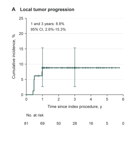
Local Tumor Progression Was 8.8% at Three Years
The primary oncological analysis included only the 81 patients whose biopsies confirmed NSCLC. Median follow-up in this subgroup was 34.5 months. Seven patients developed local tumor progression during follow-up.
All recorded local progression events occurred within the first 12 months after treatment, with a median time to progression of 5.7 months. The cumulative incidence of local progression was 8.8% at one year and remained 8.8% at three years because no additional events were recorded.
The investigators appropriately treated death without prior local progression as a competing event when estimating cumulative incidence. In a secondary Kaplan–Meier analysis, freedom from local progression was 91.2% at both one and three years.
These outcomes are encouraging. However, the absence of additional recorded progression events after one year should not be interpreted as proof that late local recurrence cannot occur.
Follow-up duration varied, and the number of patients remaining under observation decreased over time. Moreover, the study’s imaging-defined local progression endpoint differs from conventional progression-free survival, which would include disease progression outside the treated site and other relevant events.
The results therefore characterize local control in this selected cohort rather than systemic cancer control.
Three-Year Overall Survival Reached 90.8%
Overall survival in the pathology-confirmed NSCLC population was 100% at one year and 90.8% at three years. Six patients died during follow-up. The investigators reported that no deaths were attributed to the procedure or to lung cancer progression.
Among the seven patients who experienced local tumor progression, four underwent repeat cryoablation and three were referred for SBRT. However, completion of salvage treatment and subsequent outcomes were not consistently documented.
The survival results must also be interpreted in the absence of a comparator group. They cannot establish that synchronous cryoablation provides survival outcomes equivalent or superior to surgery or SBRT.
The study also did not routinely include invasive nodal staging for every patient, which introduces uncertainty regarding occult nodal disease in some cases. The findings nevertheless demonstrate that a selected group of patients with small, clinically localized NSCLC lesions experienced encouraging outcomes following the combined intervention.
Pneumothorax Remains an Important Safety Concern
The principal safety concern was pneumothorax requiring chest-tube drainage. This occurred in 21 of 102 patients, representing 20.6% of procedures. Hemorrhage or hemoptysis was recorded in five patients, while four developed clinically significant pleural effusion requiring evaluation or management.
Thirty adverse events were recorded across the study’s reported complication categories. No air embolism, needle-tract tumor seeding, or procedure-related deaths were reported. The chest-tube rate is clinically meaningful, particularly in an older population with substantial pulmonary impairment.
The investigators note that the observed rate was consistent with some published lung-ablation experiences. However, comparisons across studies are complicated by differences in patient selection, underlying lung disease, procedure complexity, and the criteria used for chest-tube placement.
A single-session strategy may reduce the need for separate procedural visits, but it does not eliminate the risks associated with percutaneous transthoracic intervention. These risks must be considered when choosing among ablation, SBRT, and other treatment options.
How Does Cryoablation Compare With Established Local Treatment?
A central question is where the findings fit into current early-stage NSCLC management. The 2025 American College of Chest Physicians clinical practice guideline continues to favor surgical resection for appropriately operable stage I NSCLC.
For patients who are not suitable candidates for surgery, the guideline suggests SBRT over percutaneous ablative procedures. However, ablation remains a reasonable option when SBRT is expected to have limited effectiveness or an unfavorable complication profile.
The new study does not provide evidence sufficient to change this treatment hierarchy. No patients were randomized to cryoablation versus SBRT, surgery, or a staged biopsy-and-ablation strategy. Therefore, the results cannot establish comparative effectiveness.
The strongest interpretation is that synchronous biopsy and cryoablation may be a feasible pathway for patients who have already been selected for ablation following multidisciplinary assessment.
A prospective comparative study would be necessary to determine whether combining the procedures reduces delays, improves patient experience, lowers healthcare resource use, or meaningfully changes clinical outcomes.
A Critical Issue: Treating Before Final Pathology
The most consequential clinical distinction is that cryoablation was performed before definitive histopathological results were available.
Although most nodules were malignant, three received specific nonmalignant diagnoses, and 13 remained indeterminate following the initial biopsy. Therefore, some patients underwent ablation without subsequently receiving a confirmed diagnosis of lung cancer.
This represents an unavoidable consideration in a synchronous treatment model. In conventional staged care, pathology may help determine whether local cancer treatment is necessary before proceeding.
In the study’s pathway, the decision to perform ablation relied on a high probability of malignancy established by imaging, smoking history, clinical assessment, and multidisciplinary review. The authors attempted to minimize inappropriate treatment by applying strict selection criteria.
However, the study does not quantify whether this approach produces a favorable balance of overtreatment risk, patient convenience, and clinical benefit compared with staged care. This question deserves particular attention in future research.
What Are the Main Limitations?
Several limitations prevent broad generalization. First, the study was retrospective and conducted at a single institution. All procedures were performed by one highly experienced interventional radiologist, potentially limiting reproducibility in centers with lower procedural volumes.
Second, there was no concurrent comparator group. The study therefore cannot establish whether the combined approach improves safety or outcomes compared with biopsy and ablation performed separately.
Third, the treatment population was highly selected, consisting of patients with small suspicious nodules, substantial smoking histories, and specific radiological characteristics. Fourth, treatment was performed before final pathology, and the NSCLC-specific efficacy analysis excluded patients with non-NSCLC or indeterminate results.
Fifth, technical success relied on visible CT-defined ablation coverage rather than consistent quantitative three-dimensional margin assessment or pathological confirmation. Finally, the local progression and survival estimates become less precise as the number of patients remaining under follow-up decreases.
These limitations reinforce the need to interpret the results as evidence of feasibility and encouraging clinical outcomes rather than definitive proof of a superior treatment strategy.
OncoDaily Takeaway
The study by Rehman and colleagues demonstrates that CT-guided core biopsy and cryoablation can be integrated into a single procedure for carefully selected patients with highly suspicious 1–2 cm pulmonary nodules.
The combined approach achieved 100% intraprocedural technical success, an 87.3% specific diagnostic yield, and 97.1% tissue adequacy for intended molecular testing. Among 81 patients with pathology-confirmed NSCLC, three-year recorded local progression was 8.8%, and overall survival reached 90.8%.
These outcomes support the feasibility of the approach. However, pneumothorax requiring chest-tube drainage occurred in approximately one in five patients, emphasizing the importance of procedural risk assessment.
Furthermore, the treatment decision preceded final pathology, meaning a proportion of patients underwent ablation without confirmed malignancy.
The study’s clinical contribution is the demonstration that diagnosis and local treatment can be combined without necessarily sacrificing tissue acquisition or procedural feasibility, not proof that this strategy should replace established treatment pathways.
For carefully selected patients who are already appropriate candidates for ablation, a single-session workflow may offer practical advantages. Prospective comparative studies are now needed to determine whether those potential advantages translate into measurable improvements in patient-centered and oncological outcomes.
Sources
- Rehman S, Naidu S, Akbarullah S, Schein J, Ramis P, Mehdi R, Suh R. Synchronous Percutaneous Biopsy and Cryoablation for Suspicious Solitary Pulmonary Nodules. Journal of Vascular and Interventional Radiology. Published online October 22, 2026. Article 109150. DOI: 10.1016/j.jvir.2026.109150
- Howington J, Souter LH, Arenberg D, et al. Management of Patients With Early-Stage Non-Small Cell Lung Cancer: An American College of Chest Physicians Clinical Practice Guideline. Chest. 2025;168:810–827. DOI: 10.1016/j.chest.2025.06.023
- Zhang T, Xu Q, Hu Y, et al. Efficacy and Safety in Synchronous Core-Needle Biopsy and Cryoablation for Highly Suspicious Malignant Pulmonary Nodule. Cancer Imaging. 2025;25:78. DOI: 10.1186/s40644-025-00901-0
- Genshaft SJ, et al. Society of Interventional Radiology Multidisciplinary Position Statement on Percutaneous Ablation of Non-Small Cell Lung Cancer and Metastatic Disease to the Lungs. Journal of Vascular and Interventional Radiology. 2021.