
A landmark multicenter study published in Nature Cancer introduces LuCaS+, a single-cell atlas of more than 2.5 million nuclei from 454 patients with non-small-cell lung cancer. By connecting malignant-cell states, immune and stromal populations, and long-term clinical outcomes, researchers identified molecular signatures associated with metastatic potential and recurrence. In stage I lung adenocarcinoma, a cell-state-derived score distinguished patients with substantially different relapse risks, suggesting that the biological composition of a tumor may provide prognostic information beyond conventional anatomical staging.
Non-small-cell lung cancer (NSCLC) encompasses a remarkably diverse collection of malignant diseases. Although patients may share the same histological diagnosis and clinical stage, their tumors can behave very differently. Some early-stage cancers recur rapidly after apparently complete surgical resection, while others remain controlled for many years.
Conventional prognostic systems rely heavily on tumor size, lymph-node involvement, distant metastasis, and other clinicopathological characteristics. Molecular testing has substantially improved treatment selection, particularly in advanced disease, but the relationship between intratumoral cellular diversity and long-term clinical outcomes remains incompletely understood.
A new study published on November 12, 2026, in Nature Cancer offers a deeper view of this complexity. In A Single-Cell Lung Cancer Atlas Reveals Cellular and Molecular Drivers of Metastasis and Clinical Outcome, Philipp Jurmeister, Philipp Keyl, Liliana Harris Mochmann, and colleagues developed LuCaS+, a multicenter atlas combining single-nucleus RNA sequencing with detailed clinical information.
The work involved investigators from German institutions in Berlin, Cologne, and Munich, with Klaus-Robert Müller, Simon Schallenberg, and Frederick Klauschen serving as joint senior authors.
The investigators identified tumor-cell populations associated with metastatic spread, characterized the molecular pathways distinguishing aggressive from more differentiated states, and examined whether integrating immune and stromal populations could improve survival prediction.
Their findings suggest that the cellular composition of an NSCLC tumor contains important prognostic information that conventional staging does not fully capture.
LuCaS+: A Single-Cell Atlas Linked to Real Clinical Outcomes
One of the major limitations of earlier single-cell cancer studies has been the availability of relatively small datasets with incomplete clinical follow-up. Although single-cell RNA sequencing can reveal extraordinary biological detail, connecting those observations to recurrence and survival requires well-characterized patient populations.
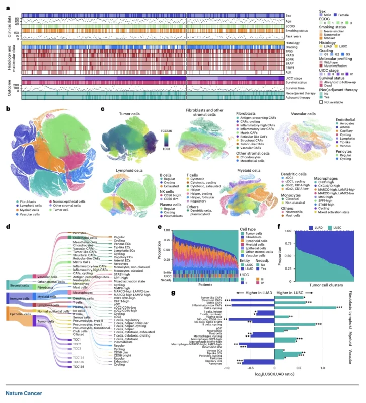
The LuCaS+ investigators addressed this challenge by analyzing archival formalin-fixed paraffin-embedded tumor specimens from three German university hospital settings. The discovery cohort comprised 364 patients treated in Berlin and Cologne. A separately processed cohort of 90 patients from Munich was used for independent validation.
Together, the atlas incorporated more than 2.5 million profiled nuclei and approximately 7.5 billion transcripts. The researchers used computational methods to identify 500 transcriptional clusters and classified 136 clusters as malignant.
They also identified 60 distinct nonmalignant cell types, including immune, stromal, vascular, and normal epithelial populations. The detailed classification included different macrophage states, T-cell populations, natural killer cells, cancer-associated fibroblasts, endothelial cells, and other microenvironmental components.
Importantly, LuCaS+ was not limited to cataloguing cell types. The investigators linked these cellular profiles to clinical characteristics, disease stage, histology, molecular alterations, and long-term follow-up. This enabled a direct investigation of whether particular tumor-cell populations were associated with metastatic disease and unfavorable survival.
Not All Cancer Cells Have the Same Metastatic Potential
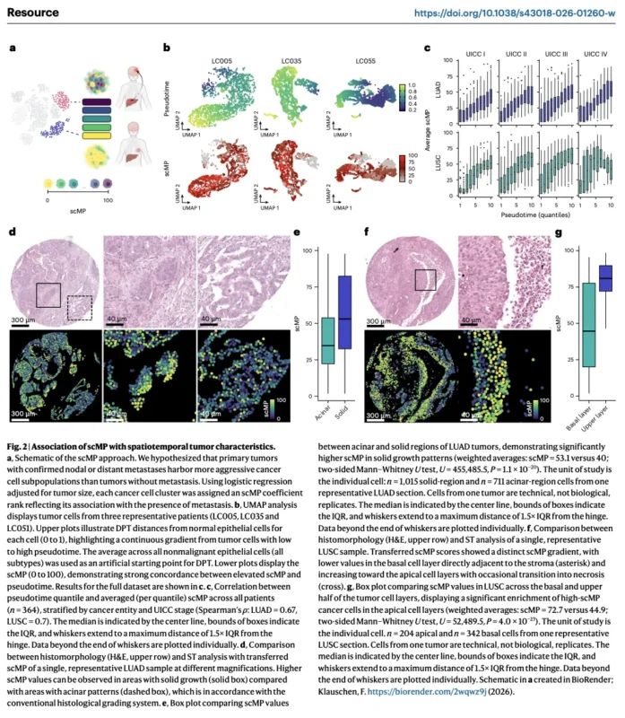
The researchers developed a metric called single-cell metastatic potential (scMP). The underlying hypothesis was that primary tumors already associated with nodal or distant metastases would contain malignant-cell populations with biological features linked to dissemination.
Using logistic regression, the investigators examined associations between the abundance of individual tumor-cell clusters and the presence of metastasis, accounting for estimated primary tumor size. Each malignant-cell cluster received an scMP score ranging from 0 to 100.
Higher values identified cell states more strongly associated with metastatic disease within the study population. This is a statistical measure derived from observational data, rather than an experimental demonstration that an individual cell can initiate metastasis.
Nevertheless, the approach revealed substantial biological differences. Cells with higher scMP scores demonstrated increased activity in pathways associated with oncogenic signaling, stress adaptation, epithelial–mesenchymal transition, and immune evasion.
The researchers also identified relationships between scMP and tumor differentiation. In lung adenocarcinoma, tumor regions showing solid growth patterns generally had higher inferred metastatic potential than areas with acinar morphology.
These findings were consistent with established histopathological observations linking certain growth patterns to aggressive disease. The analysis therefore connected microscopic tissue architecture with transcriptionally defined malignant-cell states.
Five Molecular Fingerprints of Lung Cancer Aggressiveness
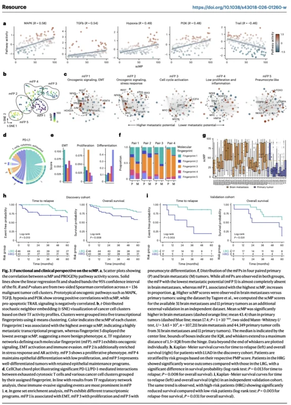
To better understand the biological basis of metastatic potential, the investigators grouped malignant-cell populations into five molecular fingerprints. The first fingerprint, designated mFP1, represented the most aggressive transcriptional state.
It was characterized by increased oncogenic signaling, epithelial–mesenchymal transition, and features associated with immune evasion. The researchers identified activity involving MYC, JUN, NF-κB, STAT1, and other regulators linked to malignant progression.
They also inferred stronger PD-L1/PD-1-associated interactions between these tumor-cell populations and cytotoxic T cells. The second fingerprint, mFP2, shared several aggressive features but showed additional stress-response and androgen receptor-associated programs.
The third fingerprint, mFP3, was dominated by cell-cycle activation and proliferation-associated regulatory networks. The fourth fingerprint represented a comparatively differentiated state with lower proliferative and inflammatory signaling.
The fifth fingerprint, mFP5, showed pneumocyte-like differentiation, including activity involving NKX2-1 and other epithelial identity regulators. Together, these fingerprints suggested a spectrum of malignant cellular states, from relatively differentiated populations to cells displaying aggressive and adaptive features.
The investigators also observed that scMP increased along computational pseudotime trajectories extending away from normal epithelial-cell states. However, pseudotime represents an inferred transcriptional ordering, not direct observation of cancer cells evolving over time.
Brain Metastases Enriched for Aggressive Cell States
The researchers examined whether their metastatic-potential framework was reflected in established metastatic lesions. They analyzed four paired primary lung tumors and corresponding brain metastases. Across these pairs, brain metastases showed increased representation of the most aggressive molecular fingerprint, mFP1.
In contrast, populations assigned to the more differentiated mFP5 state were reduced. Additional evaluation used an external dataset comprising 11 primary lung adenocarcinoma samples and 31 brain metastases from different patients.
The mean scMP score was 43.4 in brain metastases compared with 17.4 in primary tumors. These observations support an association between aggressive transcriptional states and metastatic disease. However, the analyses do not establish that these populations independently caused brain metastases.
Metastatic tumors may also acquire distinct cellular compositions through selection, adaptation, or changes in their microenvironment. Nevertheless, the findings provide an important biological link between the primary-tumor cellular landscape and metastatic progression.
A Potential New Prognostic Tool for Stage I Lung Adenocarcinoma
One of the study’s most clinically relevant findings concerned patients with stage I lung adenocarcinoma. Although surgery can provide long-term disease control for many patients, recurrence remains an important concern.
Current clinical risk assessment does not perfectly distinguish patients who will remain disease-free from those who will develop metastatic recurrence. The investigators therefore developed a patient-level measure called patient metastatic potential (PMP).
The score was calculated using the average metastatic-potential scores of the three highest-ranking malignant-cell clusters detected within a patient’s tumor. The rationale was that relatively small aggressive populations might carry meaningful prognostic information even when they did not dominate the overall tumor.
In stage I lung adenocarcinoma, PMP was associated with both relapse and overall survival. In the discovery cohort, its concordance index was 0.64 for relapse and 0.63 for overall survival. The researchers then evaluated PMP in an independent set of 26 patients with stage I lung adenocarcinoma.
In this smaller validation subset, the C-index reached 0.93 for relapse and 0.74 for overall survival. The high relapse-discrimination result is encouraging, but the limited validation sample means that the performance estimate remains uncertain and requires confirmation in substantially larger cohorts.
Major Differences in Five-Year Relapse-Free Outcomes
The investigators also explored whether PMP could divide patients into lower- and higher-risk groups. In the discovery cohort, the reported five-year relapse-free estimates were approximately 79% in the lower-risk group and 55% in the higher-risk group.
In the independent validation cohort, the corresponding estimates were 74% and 19%. In the discovery analysis, higher-risk patients had a hazard ratio of 2.85 for relapse compared with lower-risk patients. In the validation cohort, the reported relapse hazard ratio was 6.97.
These results suggest that tumor-cell states could help identify patients whose risk is underestimated by anatomical staging alone. However, the risk threshold was developed within the research framework, and the findings were not tested prospectively as a clinical decision tool.
The validation subset was small, and the observed differences should not be assumed to apply uniformly to all patients with stage I disease. The results do not establish that patients classified as high risk by PMP benefit from adjuvant chemotherapy, immunotherapy, or targeted therapy.
That question would require prospective interventional studies.
Tumor Cells Alone Do Not Explain Prognosis
A second major component of LuCaS+ focused on the tumor microenvironment. Cancer progression is influenced not only by malignant cells but also by their interactions with immune, stromal, and vascular populations.
To investigate the clinical relevance of these relationships, the researchers constructed survival-prediction models incorporating the abundance of distinct malignant and nonmalignant cell populations.
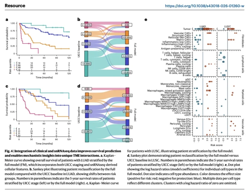
They then combined those molecular features with conventional Union for International Cancer Control (UICC) stage. In lung adenocarcinoma, the molecular model achieved a C-index of 0.70 for overall survival.
Adding anatomical stage improved the C-index to 0.727, compared with 0.650 using UICC stage alone. The improvement was statistically significant. The investigators also assessed the integrated model in a separate validation subset of 47 patients.
Here, the combined model achieved a C-index of 0.678 compared with 0.576 for UICC stage alone. These observations indicate that incorporating single-cell information may improve survival discrimination. However, the reported concordance indices reflect prediction performance in the study datasets and should not be interpreted as validated individual survival probabilities.
Stage I Patients Could Be Reclassified Into Higher-Risk Groups
The integrated model also demonstrated the ability to redistribute patients into different prognostic groups. Among 79 patients with UICC stage I lung adenocarcinoma included in the relevant analysis, 20 were reassigned to higher-risk molecular groups.
These patients had an estimated five-year survival rate of 35%, despite their early anatomical stage. Conversely, some patients with advanced-stage disease were classified into molecularly favorable groups.
Although these findings suggest that cellular composition may capture additional biological risk, the advanced-stage reassignment groups were very small. The study therefore provides evidence that molecular risk stratification may complement staging, rather than demonstrating that it should replace established clinical prognostic systems.
Lung Adenocarcinoma: Distinct Tumor–Microenvironment Interactions
The authors identified several unfavorable tumor–microenvironment phenotypes in lung adenocarcinoma. One involved TP53-defective signaling with elevated MAPK activity and reduced immune-cell infiltration.
Another represented a monocytic-inflammatory state characterized by associations among tumor cells, monocytes, and inflammatory cancer-associated fibroblasts. The investigators also identified WNT-activated, angiogenic, and TNF-associated inflammatory phenotypes.
These states differed in their inferred signaling interactions and immune context. For example, the monocytic-inflammatory phenotype showed evidence of interactions involving CSF, CXCL, FGF, and IL1/NF-κB signaling.
The authors proposed that such relationships may contribute to tumor-promoting inflammation. In contrast, other tumor niches were associated with more favorable outcomes and greater representation of cytotoxic immune populations.
These immune-permissive environments included potential interactions involving IL-2, CXCL16, and selected macrophage populations. Notably, the investigators observed that MARCO-high, LAMP2-high macrophages were associated with favorable prognosis in their dataset.
This contrasts with some previous reports associating MARCO expression with adverse outcomes and suggests that macrophage identity and function may depend on a broader combination of molecular features. These observations reinforce the limitations of treating all macrophages or fibroblasts as biologically equivalent.
Lung Squamous Cell Carcinoma: A Different Prognostic Landscape
The researchers also evaluated lung squamous cell carcinoma (LUSC). The molecular composition and prognostic relationships differed from those observed in lung adenocarcinoma. The PMP approach showed weaker and less consistent performance in stage I LUSC.
In the validation cohort, its association with relapse and overall survival was not statistically significant. This is an important limitation: a prognostic tool developed from lung cancer cell states may not perform equally across histological subtypes.
The broader integrated molecular model, however, showed greater promise. For LUSC, the combined model achieved a discovery-cohort C-index of 0.757, compared with 0.713 for UICC staging alone, although this difference had borderline statistical significance.
In independent validation, the C-index was 0.64 for the integrated model versus 0.55 for stage alone. The LUSC analysis also highlighted unfavorable microenvironments characterized by angiogenesis, matrix-associated fibroblasts, and TGFβ signaling.
VEGF–TGFβ Signaling Reveals a Potentially Self-Reinforcing Angiogenic Niche
One particularly interesting observation involved interactions among tumor cells, endothelial cells, pericytes, cancer-associated fibroblasts, and SPP1-high macrophages.
The researchers identified a molecular pattern suggesting reciprocal VEGF- and TGFβ-associated signaling. Within this inferred network, pericyte-derived TGFβ ligands could stimulate tumor cells, while tumor- and stromal-derived signals may promote macrophage recruitment and vascular activity.
Spatial transcriptomic analyses provided additional evidence that these cell types occupied related anatomical neighborhoods within tumors. This suggests that angiogenic activity may be sustained through coordinated interactions among multiple cellular compartments.
However, the findings describe inferred signaling relationships and spatial associations.
They do not establish that blocking these pathways will improve clinical outcomes in patients identified by the signature. The authors propose that these biological patterns could inform future research into more precisely selected antiangiogenic or immune-modulating strategies.
Could the Atlas Reveal New Therapeutic Targets?
Beyond prognosis, LuCaS+ offers a platform for studying treatment vulnerabilities. The authors discuss several potential strategies arising from the observed molecular states.
Examples include investigating MAPK-directed approaches in selected TP53-defective phenotypes, targeting inflammatory signaling in monocyte-associated tumor niches, and studying TGFβ inhibition in angiogenic, immunosuppressed microenvironments.
These possibilities are hypothesis-generating. The atlas does not provide clinical evidence that patients with any specific molecular fingerprint will benefit from the corresponding targeted treatment. Likewise, it does not establish new eligibility criteria for immune checkpoint inhibitors or antiangiogenic agents.
The therapeutic significance of these cellular interactions must be tested in experimental models and prospective clinical studies.
Limitations and the Path to Clinical Translation
Despite its scale, LuCaS+ has several important limitations. The analysis was retrospective and relied on archival tumor specimens from German academic centers. Most patients contributed surgical specimens, and the study population may not fully represent patients with unresectable or metastatic disease diagnosed through small biopsies.
Although the atlas included 454 patients, individual prognostic analyses often involved substantially smaller subsets. The independent stage I adenocarcinoma relapse analysis, for example, included only 26 patients.
The cellular classifications and prognostic scores were developed using complex computational methods, and some risk thresholds were optimized using the available data. Independent validation was valuable, but it does not replace prospective, multicenter clinical qualification.
Furthermore, transcriptomic associations do not necessarily establish biological causation. The identified pathways may indicate mechanisms of metastasis or resistance, but functional experiments are needed to confirm whether modifying these states changes disease behavior.
Finally, implementation of single-nucleus profiling requires specialized laboratory and computational infrastructure.
Before routine clinical adoption, future studies must demonstrate analytical reproducibility, standardized scoring, feasible turnaround times, and incremental clinical utility beyond existing pathological and molecular assessments.
The most important unanswered question is whether treatment decisions guided by these molecular profiles can improve patient outcomes.
OncoDaily Takeaway
LuCaS+ represents an important advance in connecting lung cancer cellular biology with clinically meaningful outcomes.
By profiling more than 2.5 million nuclei from 454 patients, Jurmeister and colleagues identified malignant-cell states associated with metastatic disease and revealed distinct interactions among tumor, immune, stromal, and vascular populations.
In stage I lung adenocarcinoma, a metastatic-potential score distinguished patients with substantially different relapse risks, including a promising C-index of 0.93 in a small independent validation subset. The integration of single-cell features with conventional anatomical stage also improved overall survival prediction in the studied cohorts.
Importantly, the study suggests that prognostic information may reside in relatively small aggressive tumor-cell populations that are not fully represented by conventional histology or bulk molecular analysis. However, these models remain investigational. They are not validated for routine adjuvant treatment selection, and their clinical utility has not been established prospectively.
The central implication is that the future of lung cancer precision medicine may depend not only on identifying driver mutations, but also on understanding which malignant-cell states are present and how they interact with their surrounding microenvironment.
LuCaS+ provides a substantial research foundation for developing these approaches, while prospective clinical validation will determine whether single-cell-informed risk assessment can ultimately improve patient care.
Sources
- Jurmeister P, Keyl P, Mochmann LH, et al. A Single-Cell Lung Cancer Atlas Reveals Cellular and Molecular Drivers of Metastasis and Clinical Outcome. Nature Cancer. Published November 12, 2026. DOI: 10.1038/s43018-026-01260-w