
Small cell lung cancer remains one of the most immunologically challenging solid tumors. Although platinum–etoposide combined with PD-L1 blockade has improved outcomes in extensive-stage disease, durable responses remain limited to a minority of patients. One important explanation is the frequent suppression of major histocompatibility complex class I expression, which reduces conventional CD8-positive T-cell recognition and allows SCLC cells to evade adaptive immunity.
A new study published in Cancer Cell identifies a potentially important way around this barrier. Jin Ng, Yue You, Tina Z. Zhang, and colleagues demonstrate that γδ T cells infiltrate human SCLC, retain cytotoxic activity despite PD-1 expression, and can recognize and kill tumor cells through mechanisms that do not depend on conventional MHC-I antigen presentation. The work also links higher γδ T-cell signatures with improved outcomes after PD-L1–based therapy in analyses of IMpower133 and CASPIAN and shows experimentally that γδ T cells can be recruited by tarlatamab or activated through a BTN2A1-dependent pathway enhanced by zoledronate.
The findings remain predominantly translational and preclinical; they do not establish γδ T-cell biomarkers or zoledronate-based strategies as standards of care. But they identify an immune population that may be particularly relevant in a disease whose biology frequently disables conventional antigen presentation.
Why SCLC Is Difficult to Treat With Immunotherapy
SCLC is characterized by aggressive growth, early metastatic dissemination and rapid development of treatment resistance. Immune checkpoint inhibition has changed first-line management of extensive-stage disease, but the magnitude of benefit remains relatively modest compared with that seen in several other cancers. The immune biology of SCLC helps explain this limitation.
Many SCLC tumors show reduced expression of MHC class I, together with variable CD8-positive T-cell infiltration and low tumor-cell PD-L1 expression. Because conventional αβ T cells depend heavily on antigen presentation through MHC molecules, suppression of this pathway can substantially weaken adaptive antitumor immunity. γδ T cells are biologically different.
They occupy an intermediate position between innate and adaptive immunity and can recognize cellular stress through T-cell and natural-killer-like receptors without requiring classical peptide presentation through MHC-I. Their presence in epithelial tissues, including the lung, and their relative resistance to conventional T-cell exhaustion make them particularly interesting in MHC-I-low tumors.
The investigators therefore asked whether γδ T cells might represent an overlooked immune effector population in SCLC.
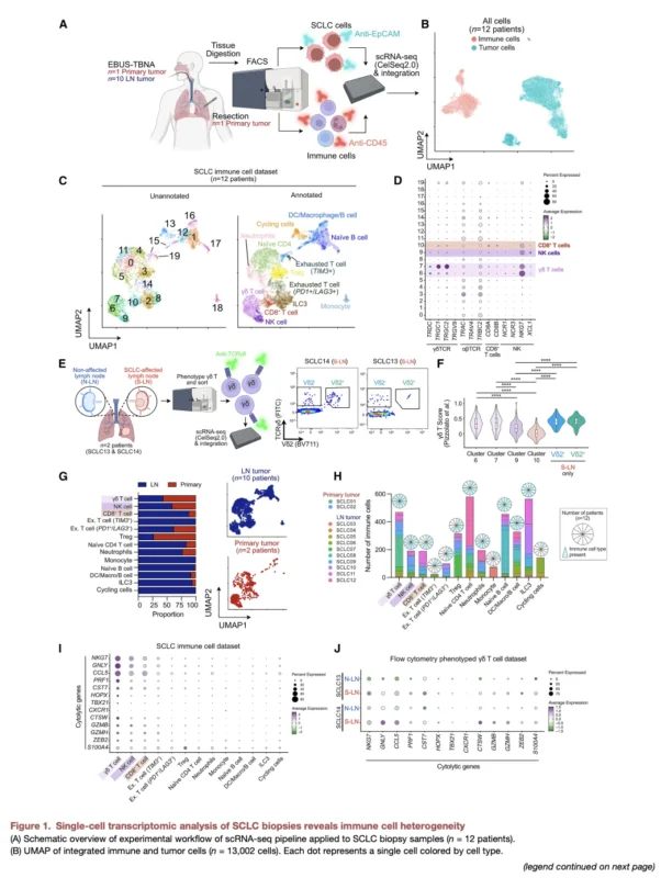
Single-Cell Profiling Identified Cytotoxic γδ T Cells Within SCLC
The study began with single-cell RNA sequencing of SCLC biospecimens from 12 patients, primarily obtained through endobronchial ultrasound-guided lymph-node sampling. After quality control, the investigators analyzed more than 13,000 single cells and identified distinct immune populations, including CD8-positive T cells, natural killer cells and γδ T cells. γδ T cells were found across all 12 patient samples and at both lymph-node and primary-tumor sites.
Their transcriptional phenotype was particularly important. γδ T cells expressed high levels of canonical cytotoxic mediators, and cells isolated from SCLC-involved lymph nodes showed stronger expression of cytolytic genes than those from nonaffected nodes. This provided evidence that the γδ population was not simply present within the tumor microenvironment but potentially equipped to participate in active tumor-cell killing.
The graphical abstract on page 1 summarizes this concept: γδ T-cell infiltration is linked with better anti-PD-L1 outcomes, while separate therapeutic strategies, tarlatamab and zoledronate, can redirect or enhance γδ T-cell activity against SCLC.
High γδ T-Cell Signatures Were Associated With Better Immunotherapy Outcomes
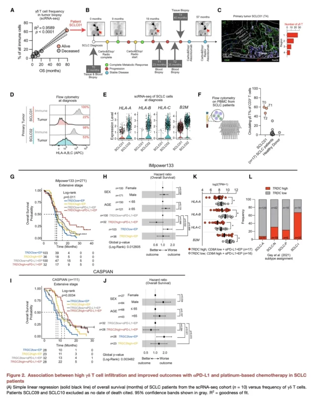
The investigators next explored whether γδ T-cell infiltration might have clinical relevance. In their initial patient cohort, a higher γδ T-cell frequency correlated with longer overall survival. One striking individual case involved a patient with unusually high γδ T-cell levels in both tumor tissue and peripheral blood who survived for more than 80 months.
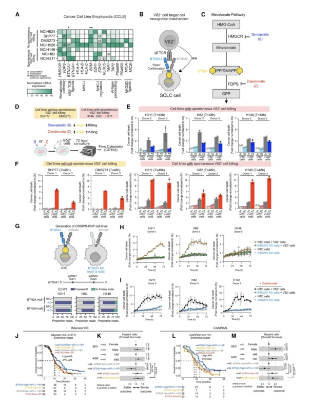
Because that small cohort could not establish a robust biomarker relationship, the investigators turned to two major randomized clinical-trial datasets. In IMpower133, involving 271 patients with extensive-stage SCLC, expression of TRDC, encoding the δ-chain of the γδ T-cell receptor, was used as a surrogate for γδ T-cell infiltration.
Among patients receiving atezolizumab plus platinum–etoposide, those with high TRDC expression had a median OS of 15.4 months compared with 10.8 months among TRDC-low tumors. High expression of TRGC2, encoding a γ-chain component, similarly identified patients with numerically longer survival following atezolizumab-based treatment.
The signal was independently examined in the CASPIAN dataset. Patients with TRGC2-high tumors receiving durvalumab plus platinum–etoposide had a median OS of 12.7 months compared with 8.6 months among TRGC2-low tumors.
These analyses are important but should be interpreted carefully. The subgroup associations were retrospective, and several reported comparisons were trends rather than independently powered prospective biomarker analyses. The study therefore supports γδ T-cell infiltration as a candidate predictive biomarker of benefit from PD-L1 blockade, not a validated test ready for clinical selection.
The Signal May Be Predictive Rather Than Simply Prognostic
A particularly relevant observation was what happened outside the immunotherapy setting. High TRDC expression was not associated with significantly improved survival in a separate limited-stage SCLC population that had not received immune checkpoint blockade.
That finding suggests the γδ T-cell signal may not simply identify patients with inherently favorable biology. Instead, it raises the possibility that these cells specifically contribute to the effectiveness of PD-L1–directed therapy. This distinction is critical.
A prognostic biomarker tells us how a patient’s disease is likely to behave irrespective of treatment. A predictive biomarker identifies patients more or less likely to benefit from a particular therapeutic strategy. The data are not sufficient to establish TRDC or TRGC2 as clinically validated predictive markers, but they provide the biological rationale to test that hypothesis prospectively.
γδ T Cells May Expand the Concept of the “Inflamed” SCLC Subtype
Previous molecular studies have identified an immune-inflamed SCLC phenotype, generally referred to as SCLC-I, that appears more responsive to checkpoint blockade. However, current immune signatures primarily emphasize conventional T-cell biology and do not specifically include γδ T-cell genes such as TRDC and TRGC2.
The current study found γδ T-cell infiltration not only in immune-inflamed disease but also across SCLC-A, SCLC-N and SCLC-P molecular phenotypes, which are typically less immunologically inflamed and often MHC-I low. This raises an important precision-oncology possibility. Some tumors currently classified as poorly immunogenic according to conventional αβ T-cell signatures may still contain a therapeutically relevant γδ T-cell compartment.
Adding γδ-associated genes to future immune classifiers could therefore potentially identify additional patients likely to benefit from immune-based treatment. That concept requires prospective validation before it can influence treatment selection.
PD-1 Expression Did Not Mean γδ T-Cell Exhaustion
Another interesting finding relates to the interpretation of PD-1. In conventional CD8-positive T cells, persistent PD-1 expression is commonly associated with a dysfunctional or exhausted phenotype. The γδ compartment behaved differently.
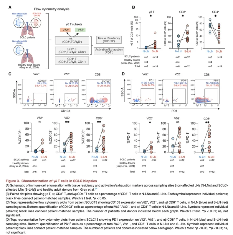
Within SCLC-involved lymph nodes, PD-1-positive γδ T cells retained cytotoxic transcriptional programs despite expressing the checkpoint receptor. The investigators also identified increased tissue-associated markers such as CD103, suggesting that these cells were activated and positioned within the tumor microenvironment rather than simply circulating bystanders.
This may help explain why γδ T cells remain functionally relevant even in tumors in which conventional antigen presentation and αβ T-cell activity are compromised.
Tarlatamab Can Recruit γδ T Cells to Kill SCLC
The study then connected these observations to one of the most important recent advances in SCLC: tarlatamab. Tarlatamab is a DLL3×CD3 bispecific T-cell engager that physically links DLL3-expressing SCLC cells to CD3-positive T cells, generating an immune synapse and promoting tumor-cell killing without requiring MHC-I antigen presentation.
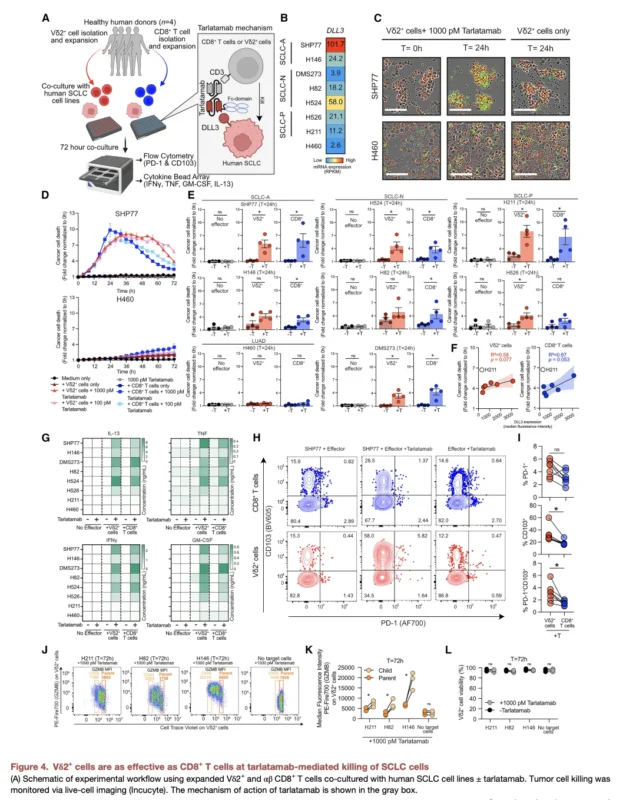
Previous work focused primarily on conventional CD4-positive and CD8-positive T cells. Ng and colleagues asked whether tarlatamab could also engage γδ T cells. Using expanded human Vδ2-positive γδ T cells, the most common circulating γδ subset, investigators co-cultured the cells with a panel of SCLC cell lines representing several molecular subtypes.
The results were notable: Vδ2-positive cells were approximately as effective as conventional CD8-positive T cells at tarlatamab-mediated SCLC killing. The detailed experiments presented in Figure 4 on pages 8–9 demonstrate tumor-cell killing across multiple SCLC models, along with γδ T-cell activation, proliferation, granzyme B induction and cytokine release after tarlatamab exposure.
This provides a new mechanistic perspective on how DLL3-directed T-cell engagement may work. Tarlatamab may be recruiting a broader immune-cell repertoire than conventional αβ T cells alone.
Tarlatamab-Mediated Killing Extended Across SCLC Molecular Subtypes
The investigators evaluated cell lines representing ASCL1, NEUROD1 and POU2F3-defined SCLC phenotypes with varying levels of DLL3. Tarlatamab promoted Vδ2-mediated killing across multiple molecular subtypes, and the magnitude of killing was generally related to DLL3 surface expression.
The broader point is that γδ T cells provide a functional effector population downstream of CD3 engagement. Rather than requiring endogenous tumor-specific antigen recognition, tarlatamab brings the γδ T cell directly to the DLL3-positive tumor cell and activates cytotoxicity.
This MHC-independent mechanism is particularly attractive in SCLC because MHC-I loss is one of the tumor’s characteristic mechanisms of immune escape.
Zoledronate Revealed a Second, Independent γδ T-Cell Strategy
Perhaps the most unexpected element of the study was the finding involving zoledronate. Vδ2-positive γδ T cells can recognize intracellular metabolic stress through phosphoantigens generated within the mevalonate pathway. This recognition involves interaction between the Vδ2 T-cell receptor and BTN2A1/BTN3A1 complexes on target cells.
Zoledronate inhibits farnesyl pyrophosphate synthase, leading to intracellular accumulation of phosphoantigens. In the study, zoledronate increased Vδ2-positive T-cell killing of SCLC lines that were already susceptible and, importantly, sensitized previously resistant SCLC cells to γδ-mediated killing.
This was mechanistically distinct from tarlatamab. Tarlatamab creates an artificial DLL3–CD3 bridge. Zoledronate increases tumor-cell visibility to the intrinsic Vδ2 T-cell receptor system. The convergence of two very different strategies on the same immune effector population makes γδ T cells especially interesting therapeutically.
BTN2A1 Was Required for MHC-Independent Recognition
The investigators directly interrogated the molecular mechanism using CRISPR-mediated knockout of BTN2A1 in SCLC cells. Loss of BTN2A1 substantially reduced Vδ2-positive T-cell recognition and tumor-cell killing, both under baseline conditions and following zoledronate exposure. This establishes BTN2A1 as a critical component of the recognition axis in these experimental systems. The clinical datasets provided another intriguing observation.
In IMpower133, high BTN2A1 expression was associated with longer OS among patients receiving atezolizumab plus platinum–etoposide: 14.4 months versus approximately 9.7–10.4 months in the other groups, with an HR of 0.67. The association was also reproduced directionally in CASPIAN, where the BTN2A1-high group receiving durvalumab plus chemotherapy had the greatest reported survival.
These retrospective analyses again require validation, but together with the experimental data they provide a coherent biological story: BTN2A1 may represent an MHC-independent gateway through which Vδ2 γδ T cells recognize SCLC.
Patient-Derived Organoids Strengthen the Translational Evidence
The investigators did not restrict their experiments to established cancer cell lines. They generated SCLC organoids from diagnostic biopsies and simultaneously expanded Vδ2-positive cells from peripheral blood. In one patient, zoledronate plus IL-2 expanded Vδ2-positive cells approximately 260-fold over 13 days. Similar expansion was demonstrated in additional patients.
Patient-matched Vδ2-enriched immune cells increased killing of autologous SCLC organoids when combined with either zoledronate or tarlatamab. The effect was also reproduced in allogeneic models using γδ T cells from healthy donors.
These experiments remain ex vivo, but they strengthen the biological plausibility of γδ-directed strategies beyond simplified tumor-cell-line models.
Zoledronate Should Not Yet Be Viewed as an SCLC Treatment
The repurposing concept is attractive because zoledronate is already widely used in oncology. However, the study does not demonstrate that administering zoledronate to patients with SCLC improves immunotherapy response or survival. Its pharmacology also creates an important challenge: zoledronate has strong affinity for bone, which may limit exposure to extracranial SCLC tumors.
The authors note that this issue requires further investigation and suggest that targeted delivery approaches, including DLL3-directed platforms, could potentially enhance delivery to SCLC cells. The zoledronate findings should therefore be viewed as a mechanistic proof of concept for manipulating the Vδ2–BTN2A1 pathway, rather than an immediate repurposing recommendation.
Could γδ T Cells Become a Biomarker for Checkpoint Therapy?
The retrospective IMpower133 and CASPIAN analyses create a clinically important hypothesis. Unlike NSCLC, SCLC still lacks a robust routine biomarker for selecting patients most likely to benefit from PD-L1 inhibition. PD-L1 expression itself has limited utility, and tumor mutational burden has not become a standard selection tool. γδ-associated signatures may provide a different way to assess the tumor immune microenvironment.
Because γδ T cells can remain functional even in MHC-I-low disease, they may identify a subset of tumors with meaningful immune vulnerability that is invisible to conventional CD8-focused signatures. But several steps are necessary before this becomes clinically useful: standardized methods for quantifying γδ T cells, prespecified thresholds, prospective clinical validation and demonstration that the biomarker truly predicts differential treatment benefit.
At present, TRDC, TRGC2 and BTN2A1 remain research biomarkers.
The Study Suggests Multiple Therapeutic Routes Through the Same Immune Cell
One of the strongest aspects of the paper is that it does not identify only one therapeutic mechanism. It outlines several potential ways to exploit γδ biology:
- checkpoint therapy, in tumors already containing active γδ T cells
- tarlatamab, which can redirect γδ T cells to DLL3-positive cancer cells
- zoledronate or related metabolic manipulation, which can increase Vδ2 recognition through the BTN2A1 pathway
- potentially adoptive γδ T-cell therapy, using ex vivo expanded effector populations
These strategies are mechanistically distinct but converge on an immune-cell population capable of bypassing one of SCLC’s fundamental resistance mechanisms: loss of classical antigen presentation. That makes γδ T cells less a single drug target than a potential immunologic platform.
Important Limitations Keep the Findings Translational
The paper combines human biopsy profiling, retrospective clinical-trial datasets, cancer cell lines, genetic experiments and patient-derived organoids, providing unusually broad mechanistic support. Nevertheless, most therapeutic conclusions remain preclinical.
The initial single-cell SCLC cohort included only 12 patients. The associations with IMpower133 and CASPIAN were retrospective biomarker analyses rather than prospectively stratified treatment comparisons. Some subgroup hazard ratios had broad confidence intervals and were described as trends.
Tarlatamab-mediated γδ killing was demonstrated primarily in vitro rather than proven as the dominant mechanism in treated patients. The zoledronate strategy likewise has not undergone prospective SCLC clinical testing. The remarkable long survival of the individual patient with exceptionally high γδ T-cell levels is biologically intriguing but cannot establish causation.
Accordingly, the study should be interpreted as defining a new immune vulnerability and therapeutic hypothesis, not a new treatment standard.
The Bottom Line
The Cancer Cell study by Ng and colleagues identifies γδ T cells as an underrecognized component of antitumor immunity in SCLC. These cells were detected throughout human SCLC samples and retained strong cytotoxic programs despite PD-1 expression. Higher γδ-associated transcriptional signatures were linked with improved outcomes after atezolizumab or durvalumab-based therapy in retrospective analyses of IMpower133 and CASPIAN.
Experimentally, Vδ2-positive γδ T cells were as effective as conventional CD8-positive T cells in tarlatamab-mediated SCLC killing, demonstrating that DLL3-directed T-cell engagement may recruit a broader immune repertoire than previously appreciated.
A second pathway emerged through BTN2A1, which enabled MHC-independent Vδ2 recognition of SCLC. Zoledronate increased phosphoantigen accumulation and sensitized SCLC cells to γδ-mediated killing, while BTN2A1 knockout substantially attenuated this activity. The broader significance is conceptual. SCLC has traditionally been viewed as an immunologically difficult tumor partly because it suppresses the machinery needed for conventional T-cell recognition. γδ T cells provide a potential way around that problem.
Rather than attempting only to restore classical antigen presentation, future immunotherapy strategies may exploit immune cells that never required MHC-I recognition in the first place.
That possibility, combined with checkpoint blockade, DLL3 engagement, metabolic sensitization or cellular therapy, opens a new translational direction for SCLC immunotherapy.
Reference
- Ng J, You Y, Zhang TZ, Hess JB, Best SA, Caneborg A, Schmiel M, Diepstraten ST, Spelman T, Gray JI, Godfrey DI, Wu Y, Tothill RW, Anttila CJA, Baldwin TM, Naik SH, Amann-Zalcenstein D, Kersbergen AJ, Leong TL, George J, Ritchie ME, Gherardin NA, Koay H-F, Hickey PF, Steinfort D, Sutherland KD. γδ T cells modulate anti-tumor immunity in small cell lung cancer. Cancer Cell. 2026;44:1–18. doi:10.1016/j.ccell.2026.08.015.