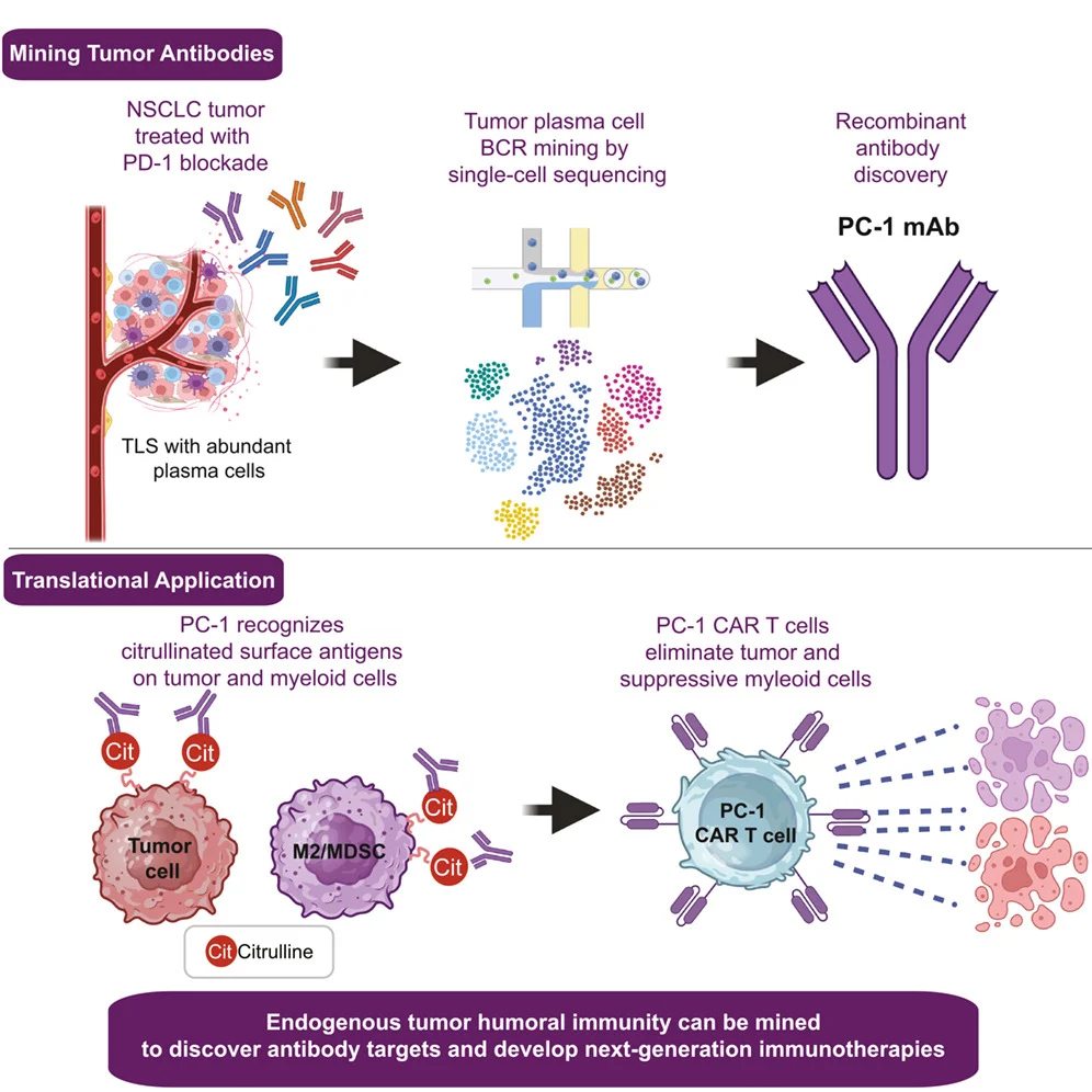
A new study published in Cancer Cell suggests that the humoral immune response within tumors after PD-1 blockade may provide more than a marker of antitumor immunity—it may also offer a source for discovering new cancer therapeutics. In “Tumor-infiltrating plasma cell profiling after PD-1 blockade reveals tumor-specific antibodies,” R. Ryan Meyerhoff and colleagues analyzed tumor-infiltrating B-cell populations from patients with non-small cell lung cancer (NSCLC) treated with neoadjuvant PD-1 blockade. By combining spatial profiling, single-cell transcriptomics, B-cell receptor (BCR) sequencing, and functional antibody testing, the investigators identified tumor-reactive antibody lineages within the tumor microenvironment.
One antibody, termed PC-1, emerged as the lead candidate. PC-1 recognized tumor-associated citrullinated proteins and bound not only malignant cells but also several tumor-promoting populations within the microenvironment. The investigators subsequently converted its antigen-recognition domain into a chimeric antigen receptor (CAR), generating PC-1 CAR T cells that demonstrated antitumor activity in preclinical models. The findings raise a broader immuno-oncology question: could tumor-infiltrating B-cell responses provide a source of naturally selected antibodies that can be repurposed into new cancer therapeutics?
Beyond T Cells: What Happens to B Cells After PD-1 Blockade?
The conventional biological framework for PD-1 blockade has centered largely on T cells, with checkpoint inhibition restoring or sustaining the activity of tumor-reactive T cells constrained within the tumor microenvironment. Increasing evidence, however, suggests that B cells also contribute to antitumor immunity through antigen presentation, regulation of T-cell function, and antibody production. Tumor-infiltrating B cells and their organization within tertiary lymphoid structures (TLSs) have been associated with favorable outcomes across several malignancies, while previous NSCLC studies have linked TLS abundance and intratumoral plasma cells with response and clinical outcomes following checkpoint-based treatment.
Meyerhoff and colleagues moved beyond measuring the abundance or phenotype of these cells and instead asked what their antibodies actually recognize. By reconstructing BCR sequences from tumor-infiltrating B-cell populations and producing selected antibodies for functional testing, the investigators examined whether the humoral immune response present within PD-1-treated tumors contained antibodies capable of recognizing cancer-associated surface antigens. This shifts the focus from B cells as markers of an immune-active tumor toward B cells as potential sources of therapeutic antigen recognition.
Mining the B-Cell Repertoire of PD-1-Treated NSCLC
The human tumor specimens originated from the previously reported phase II TOP1501 trial (NCT02818920), a single-arm study evaluating neoadjuvant pembrolizumab in patients with clinical stage IB, II, or IIIA NSCLC before surgical resection. Patients received two cycles of pembrolizumab 200 mg intravenously before surgery. For the current translational analysis, the investigators identified five post-resection tumor specimens with tumor-infiltrating B cells and sufficient banked material for further study, underscoring the relatively small size of the human discovery cohort.
Spatial profiling of available resection specimens demonstrated numerous TLSs, including mature structures containing germinal-center CD4+ T cells, proliferating B cells, and CD138+CD38+ antibody-secreting cells. Plasma cells were also distributed throughout the tumor stroma and parenchyma, supporting the presence of a local environment capable of B-cell maturation and antibody production. The investigators did not find a clear association between TLS maturation state and pathological response in these specimens, which they noted could reflect the limited tissue available and sampling variability.
Single-cell transcriptomic profiling subsequently identified 7,150 quality-filtered tumor-infiltrating B cells across several states, including naive-like, activated naive, cycling, germinal-center, memory, atypical-memory, and antibody-secreting populations. Integration with reconstructed BCR sequences yielded 5,281 functional paired heavy- and light-chain sequences. Germinal-center, memory, and antibody-secreting populations demonstrated class switching and greater somatic hypermutation than naive-like B cells, while shared clonal lineages across B-cell states provided evidence of differentiation and clonal expansion within NSCLC tumors.
Were the Intratumoral Antibodies Actually Tumor-Reactive?
To determine whether these B-cell populations contained antibodies capable of recognizing tumor-associated surface antigens, the investigators generated recombinant monoclonal antibodies from selected BCR sequences. Among 15 antibodies representing some of the most expanded tumor-infiltrating plasma-cell clonotypes, 11 of 15 (73%)demonstrated cell-surface antigen reactivity against the Calu-6 NSCLC cell line. The investigators then broadened the analysis by generating 29 additional antibodies from germinal-center, memory, atypical-memory, and less clonally expanded but highly mutated or diversified antibody-secreting lineages. Of these, 15 of 29 (52%) showed cell-surface reactivity against A549 NSCLC cells.
These findings indicate that tumor-reactive antibody specificities were present across several antigen-experienced intratumoral B-cell states rather than being restricted to the most expanded plasma-cell populations. Among the antibodies tested, PC-1 showed particularly strong cell-surface reactivity and represented one of the most clonally expanded antibody-secreting cell lineages, with 107 recovered members. Its combination of clonal expansion and tumor reactivity led the investigators to select PC-1 for more detailed functional and antigen-specificity studies.
PC-1 Reveals Citrullinated Proteins as Tumor-Associated Targets
PC-1 bound surface antigens across cancer cell lines derived from several anatomical sites, including lung, mesothelium, ovary, and uterus, suggesting that its target was not restricted to a single tumor lineage. Immunoprecipitation followed by mass spectrometry identified vimentin as a leading candidate antigen. Although vimentin is normally an intracellular structural protein, it can appear at the cell surface in a citrullinated form, a post-translational modification that has been described as a potential source of tumor-associated antigens.
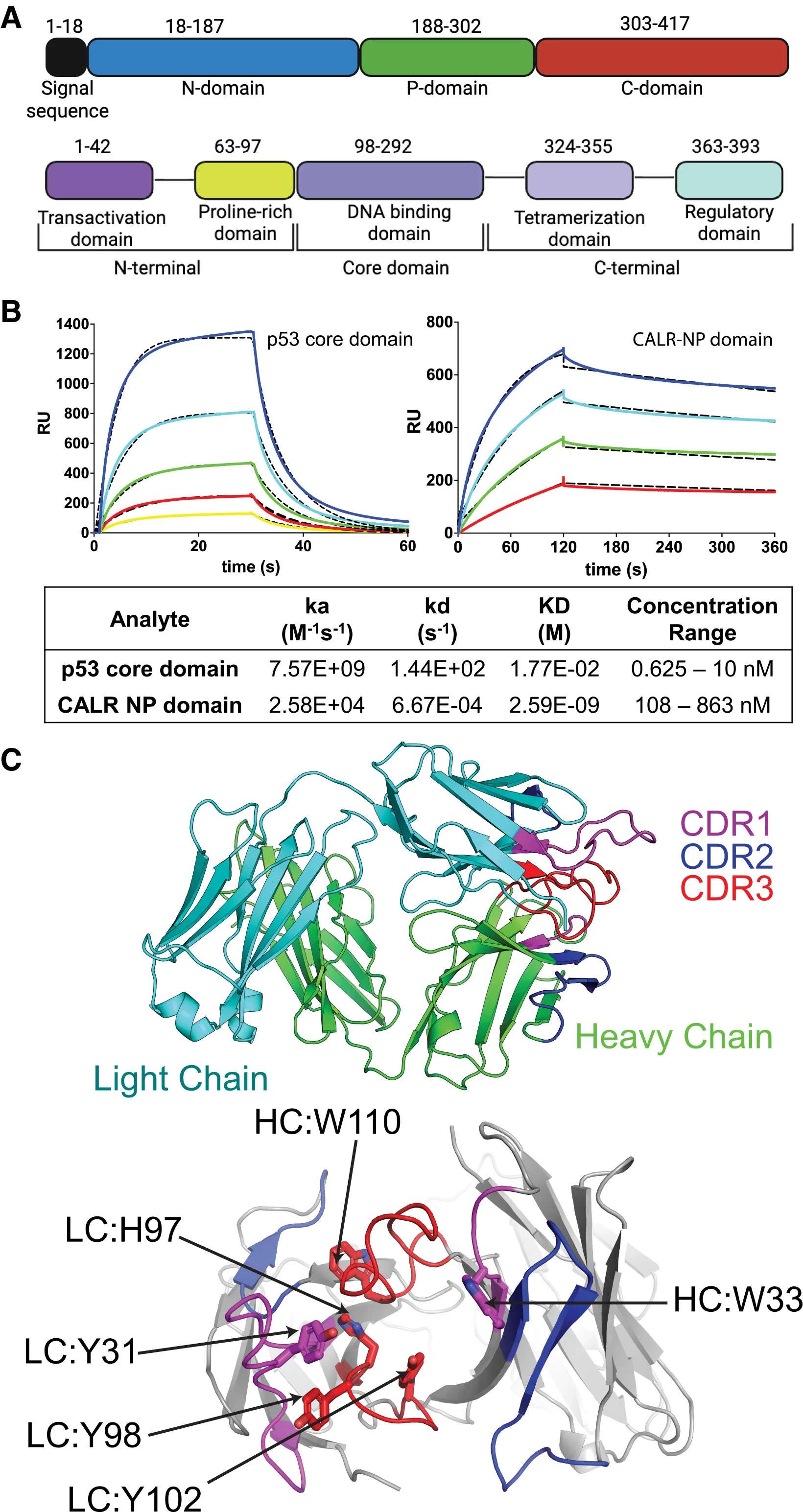
PC-1 bound recombinant citrullinated vimentin but not unmodified vimentin and also demonstrated high-affinity binding to other proteins, including calreticulin and p53. Several additional tumor-derived antibodies showed reactivity against citrullinated calreticulin, suggesting that recognition of post-translationally modified proteins may extend beyond the PC-1 lineage. The precise structural basis of PC-1 recognition remains unresolved, however, and the authors propose that citrullination may expose or stabilize shared conformational features rather than citrulline alone serving as the antibody’s binding determinant.
This biology is particularly interesting within the tumor microenvironment. Citrullination can occur during inflammation, cellular stress, tissue injury, myeloid activation, and immunogenic cell death. Whereas these processes are generally transient in normal tissues, tumors provide a chronic environment characterized by hypoxia, cell death, inflammatory signaling, and tissue remodeling. The authors suggest that this context could contribute to abnormal surface exposure of modified intracellular proteins and create antigenic states that distinguish malignant or tumor-associated cells from their normal counterparts.
Does PC-1 Represent Pathological Autoreactivity?
Recognition of citrullinated proteins raises an important question because antibodies against citrullinated antigens are associated with autoimmune disorders, particularly rheumatoid arthritis. The investigators therefore examined whether PC-1 shared characteristics with conventional pathological autoantibodies. PC-1 did not bind the cyclic citrullinated peptides used in their analysis and did not demonstrate reactivity against a clinical panel of nuclear autoantigens associated with systemic autoimmune diseases. The patient from whom PC-1 was isolated had no clinical manifestations of rheumatoid arthritis before or after checkpoint blockade.
Additional profiling nevertheless illustrates why clinical safety cannot be inferred from these experiments alone. PC-1 demonstrated intense cell-surface binding to thyroid epithelial cells in a normal-tissue microarray, although binding across other tissues varied, and testing against 23,004 proteins and isoform variants showed above-cutoff binding to only 0.6% of proteins. The antibody therefore did not show the conventional autoreactivity pattern examined by the investigators, but broader tissue recognition remains relevant when considering future therapeutic development.
Targeting the Tumor and Its Immunosuppressive Microenvironment
One of the most interesting observations came from testing PC-1 against patient-derived tumor material. The antibody bound freshly dissociated high-grade serous ovarian carcinoma cells but not paired normal tissue. It also recognized several tumor-associated populations, including tumor-promoting macrophages, myeloid-derived suppressor cells, and exhausted CD4+ T cells in ovarian tumors, as well as tumor-associated cells including cancer-associated fibroblasts in NSCLC malignant effusions. In contrast, PC-1 did not react with immune cells from healthy-donor peripheral blood.
This pattern suggests a potentially interesting therapeutic property because the recognized antigenic state may extend beyond malignant cells to components of the microenvironment that support tumor progression or immune suppression. Solid-tumor therapies frequently face both tumor-cell heterogeneity and a microenvironment that limits effective immune-cell function. A recognition system capable of targeting malignant cells together with selected tumor-promoting populations could therefore have biological advantages, although the current experiments provide only preclinical proof of concept and do not establish that such dual targeting will translate into improved clinical efficacy.
From a Tumor-Derived Antibody to CAR T Cells
The investigators next tested whether an antibody specificity discovered from an endogenous intratumoral B-cell response could be converted into an engineered cellular therapy. They incorporated the PC-1-derived single-chain variable fragment into CAR constructs containing either CD28 or 4-1BB costimulatory domains. Both PC-1 CAR T-cell designs demonstrated cytotoxicity against NSCLC cell lines in vitro, and CD28-costimulated PC-1 CAR T cells controlled established Calu-6 and A427 tumor growth in xenograft models compared with mock-transduced T cells.
To test whether this activity depended on protein citrullination, the researchers generated PAD2-deficient A427 tumor cells. Loss of PAD2 reduced PC-1 surface binding from 82.6% in parental cells to 6.4% in PAD2-deficient cells and eliminated killing by both CD28- and 4-1BB-costimulated PC-1 CAR T cells. These experiments provide functional evidence that PAD2-dependent citrullinated cell-surface antigens were required for PC-1-mediated recognition and cytotoxicity rather than merely being associated with antibody binding.
The researchers extended these experiments into an immunocompetent setting using a murinized version of the PC-1 CAR. Murinized PC-1 CAR T cells killed mouse tumor cells in vitro and improved survival compared with mock-transduced T cells in an orthotopic syngeneic tumor model, despite treatment being administered without lymphodepletion. Histological examination of the lung, heart, liver, kidney, and brain showed no detectable treatment-associated tissue injury, and serum markers of renal, pancreatic, and hepatic function were not significantly different between groups. Tumors from PC-1 CAR T-treated mice, in contrast, demonstrated extensive tissue injury. Together, these experiments strengthen the preclinical evidence that the PC-1 specificity can be translated into a functional CAR platform, while remaining insufficient to establish either safety or efficacy in humans.
Could the Tumor’s Own Immune Response Become a Drug-Discovery Platform?
The broader significance of the study extends beyond PC-1 or citrullinated vimentin. The investigators effectively used the adaptive immune response within the tumor as a discovery platform: tumor-infiltrating B cells were characterized at single-cell resolution, their BCR sequences were reconstructed, selected antibodies were produced and screened for tumor reactivity, and one naturally occurring tumor-reactive antibody specificity was subsequently incorporated into an engineered CAR T-cell platform.
This creates a potentially different path for therapeutic discovery. Traditional approaches frequently begin by selecting a candidate tumor antigen and then engineering an antibody or cellular therapy against it. Mining intratumoral B-cell repertoires reverses part of that process by asking which tumor-associated structures the patient’s own humoral immune response has already selected and affinity-matured to recognize. The authors suggest that antibodies and antigens discovered through this approach could potentially contribute not only to CAR T-cell development but also to antibody-drug conjugates and future cancer vaccine platforms.
The possibility is particularly relevant for solid tumors, where identifying surface antigens with sufficient tumor selectivity remains one of the major obstacles to engineered cellular therapy. An endogenous antibody response does not automatically guarantee a safe therapeutic target, but it may provide a biologically informed starting point for identifying antigenic states enriched within malignant tissue.
Does PD-1 Blockade Generate These Tumor-Reactive Antibodies?
The relationship between checkpoint inhibition and the antibody responses observed in this study requires careful interpretation. The analyzed tumors were obtained after neoadjuvant pembrolizumab, and the investigators observed mature TLSs, plasma-cell accumulation, class-switched B-cell populations, somatic hypermutation, and clonally expanded tumor-reactive antibody lineages. These findings are consistent with an active intratumoral humoral immune response in the setting of PD-1 blockade.
The study does not, however, demonstrate that pembrolizumab generated the PC-1 lineage or that PD-1 blockade was required for its development. Systematic paired pre-treatment and post-treatment tissue was not available across the analyzed patients, and plasma antibody binding to the examined citrullinated antigens did not significantly increase after pembrolizumab. There was also no correlation between circulating antibody binding to these antigens and pathological response, leading the investigators to suggest that at least some of these humoral responses may be localized to the tumor microenvironment.
Longitudinal studies will therefore be needed to determine whether PD-1 blockade expands pre-existing tumor-reactive B-cell clones, promotes their maturation within TLSs, changes their antigen specificity, or simply provides a tumor environment in which these responses can be more readily identified. For now, the evidence supports the presence of therapeutically interesting antibody repertoires in PD-1-treated tumors rather than a causal conclusion that checkpoint blockade created them.
Limitations
The study is primarily a mechanistic and preclinical proof-of-concept investigation, and its human component is small. Only a limited number of post-neoadjuvant NSCLC specimens contained sufficient residual tissue and plasma-cell infiltrates for detailed analysis, while tissue availability prevented systematic assessment of paired adjacent normal lung and tumor-draining lymph nodes. The authors also selected clonally expanded, highly mutated, or diversified BCR lineages for recombinant antibody production, meaning that the study did not comprehensively sample the full diversity of tumor-reactive B-cell specificities.
The cellular origin and development of the antibody-secreting populations also remain incompletely defined. The investigators could not determine whether the studied cells developed within intratumoral TLSs or migrated into the tumor from systemic circulation, and the progression of the PC-1 lineage through a germinal-center reaction could not be reconstructed. Most importantly, the therapeutic findings remain preclinical. Although PC-1 CAR T cells demonstrated antitumor activity without detectable off-tumor organ injury in the reported mouse experiments, these models cannot establish clinical safety, efficacy, or the therapeutic window of targeting PC-1-reactive antigens in humans.
You Can Read Full Article Here.