Immune checkpoint inhibitors have transformed the management of many cancers, delivering durable responses in melanoma, non-small cell lung cancer, renal cell carcinoma, hepatocellular carcinoma, urothelial cancer, and other malignancies.
Yet most patients still do not achieve long-term benefit.
Why do some tumors remain resistant—even when PD-L1 is expressed or immune cells are present? An increasingly important answer may lie in the tumor’s oxygen-deprived microenvironment.
In the review “HIF-1 and HIF-2 in Cancer: Structure, Regulation, and Therapeutic Prospects,” Yi Shi and Daniele M. Gilkes examine how hypoxia-inducible factors shape cancer biology and why HIF signaling has become a compelling therapeutic target. Complementing this work, Jiayu Liu, Ying Jiang, Lingyan Chen, Zhiwen Qian, Yan Zhang, and colleagues explore the direct relationship between HIF signaling and immune checkpoints in their review, “Associations Between HIFs and Tumor Immune Checkpoints: Mechanism and Therapy.”
Together, these articles highlight a central immuno-oncology concept: hypoxia is not simply a feature of aggressive tumors. It is an active driver of immune escape.
Hypoxia Is More Than a Metabolic Problem
As solid tumors grow beyond the capacity of their abnormal vasculature, oxygen delivery becomes limited. This leads to chronic or intermittent hypoxia and stabilization of hypoxia-inducible factors, particularly HIF-1α and HIF-2α.
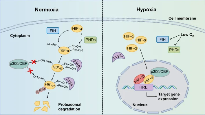
Under normal oxygen conditions, HIF-α proteins are hydroxylated and marked for degradation through the von Hippel–Lindau pathway. Under hypoxia, this process is interrupted. HIF proteins accumulate, enter the nucleus, and activate transcriptional programs that support tumor-cell survival.
These programs include angiogenesis, glycolysis, metabolic adaptation, extracellular acidification, invasion, and treatment resistance.
However, their importance extends much further. HIF signaling can reorganize the entire tumor immune microenvironment.
How Does HIF Create an Immune-Resistant Tumor?
A hypoxic tumor is often a hostile environment for effective antitumor immunity.
HIF-driven VEGF production promotes angiogenesis, but the vessels that form are typically abnormal, disorganized, and poorly perfused. This perpetuates hypoxia and makes it more difficult for activated T cells to enter the tumor.
At the same time, tumor cells shift toward glycolysis, consuming glucose and generating lactate. The resulting nutrient competition and extracellular acidification can impair the proliferation, cytokine production, and cytotoxic activity of CD8⁺ T cells and natural killer cells.
Hypoxia can also favor the accumulation or functional activity of several immunosuppressive populations, including regulatory T cells, myeloid-derived suppressor cells, and tumor-associated macrophages. Dendritic-cell maturation and antigen presentation may be weakened, limiting effective priming of tumor-specific T cells from the beginning.
The result is not simply an immune-cold tumor. It is a tumor actively protected by metabolic, vascular, and immunological barriers.
HIF and PD-L1: Why Checkpoint Blockade May Not Be Enough
The review by Liu and colleagues, “Associations Between HIFs and Tumor Immune Checkpoints: Mechanism and Therapy,” describes HIF signaling as an important upstream regulator of immune-checkpoint biology.
Preclinical studies indicate that HIF-1α can promote PD-L1 expression in tumor cells and myeloid populations. This strengthens inhibitory PD-1/PD-L1 signaling and suppresses the activity of cytotoxic T cells.
Hypoxia may also contribute to T-cell exhaustion and the simultaneous expression of several inhibitory receptors, including PD-1, TIM-3, LAG-3, and TIGIT.
This is clinically important. A tumor may express PD-L1 and still remain resistant to anti–PD-1 or anti–PD-L1 therapy because hypoxia is sustaining multiple, parallel mechanisms of immune suppression.
Adenosine: The Metabolic Checkpoint
One particularly important HIF-associated pathway is the CD39/CD73–adenosine axis.
Extracellular ATP released by stressed or dying cells can normally act as a danger signal. In hypoxic tumors, however, increased CD39 and CD73 activity converts ATP into adenosine.
Adenosine suppresses cytotoxic T-cell activation, interferon-γ production, natural killer-cell function, dendritic-cell maturation, and macrophage activation. At the same time, it supports an immunosuppressive tumor microenvironment.
This makes adenosine a metabolic checkpoint—one that may work alongside PD-1/PD-L1 signaling to limit effective antitumor immunity.
Why HIF-2α Has Become a Clinically Relevant Target
HIF-1α and HIF-2α share important functions but are not interchangeable. HIF-1α is commonly associated with acute hypoxic adaptation and glycolytic reprogramming, while HIF-2α has a particularly important role in chronic hypoxia, vascular biology, and clear-cell renal cell carcinoma.
This biology has led to the development of selective HIF-2α inhibitors, including belzutifan.
Belzutifan has validated HIF-2α as a clinically actionable target in von Hippel–Lindau disease-associated tumors and renal cell carcinoma. Its success has also raised a broader immuno-oncology question: can inhibition of HIF signaling help remodel the tumor microenvironment and improve responses to immune checkpoint blockade?
That question is now driving growing interest in rational combination strategies.
The Future May Be Combination Immunotherapy
Targeting the hypoxia–HIF axis could potentially address several resistance mechanisms at the same time:
- abnormal tumor vasculature and immune exclusion;
- PD-L1 and other checkpoint-related immune suppression;
- lactate-driven metabolic dysfunction;
- adenosine-mediated suppression;
- impaired dendritic-cell and T-cell function;
- accumulation of suppressive myeloid cells.
Potential combination approaches include HIF inhibitors with PD-1/PD-L1 blockade, VEGF-directed therapies, adenosine-pathway inhibitors, and potentially cellular immunotherapies.
Importantly, this remains an evolving field. Much of the mechanistic rationale is preclinical, and the optimal biomarkers, tumor settings, sequencing, and treatment combinations still need to be defined in prospective clinical trials.
What Does This Mean for Oncology Practice?
Hypoxia should no longer be considered only a marker of tumor aggressiveness.
It is increasingly understood as a dynamic and potentially actionable contributor to immunotherapy resistance. By linking abnormal vasculature, tumor metabolism, immune exclusion, checkpoint expression, and T-cell dysfunction, HIF signaling sits at the crossroads of several of the most important challenges in cancer immunotherapy.
The future of precision immuno-oncology may therefore depend not only on identifying the correct immune target, but also on changing the conditions within the tumor that prevent the immune system from functioning.