
The combination of targeted radiopharmaceutical therapy and immune checkpoint inhibition represents an attractive strategy in metastatic castration-resistant prostate cancer (mCRPC). Radiation delivered directly to PSMA-expressing tumor cells may produce tumor-cell death and inflammatory signaling, potentially increasing antigen availability and immune recognition in a disease in which checkpoint inhibition alone has limited activity.
Yet combining two therapeutic modalities also creates a more complex biomarker problem. If a patient responds, is the tumor intrinsically sensitive to PSMA-targeted radiation, susceptible to immune modulation, or dependent on an interaction between the two? And when resistance develops, does it emerge through loss of PSMA expression, genomic evolution, persistence of resistant clones, or several of these processes simultaneously?
A September 2026 study by Tolmeijer et al. in the Journal of Nuclear Medicine examined these questions through an integrated translational analysis of the phase 1b/2 PRINCE trial, combining longitudinal circulating tumor DNA (ctDNA), circulating tumor cells (CTCs), genomic profiling, PSMA PET and ^18F-FDG PET in patients treated with ^177Lu-PSMA-617 plus pembrolizumab.
Rather than identifying a single response biomarker, the study reveals how different measurements describe distinct dimensions of tumor biology and, importantly, how molecular and imaging responses can diverge during therapy.
Radiopharmaceutical Therapy and Immune Checkpoint Inhibition
PSMA-targeted radioligand therapy has established clinical activity in PSMA-positive mCRPC, but PSMA expression alone does not fully explain therapeutic sensitivity. Even after imaging-based patient selection, a clinically important proportion of patients derive limited benefit from ^177Lu-PSMA-617.
The rationale for adding immune checkpoint inhibition is supported by preclinical evidence that radiation from ^177Lu-PSMA-617 can promote immunogenic cancer-cell death, releasing tumor antigens and inflammatory signals that may facilitate immune recognition. This provides a potential biological basis for combining radiopharmaceutical therapy with PD-1 blockade in mCRPC, where checkpoint inhibitor monotherapy has relatively limited activity in unselected disease.
In PRINCE, patients received ^177Lu-PSMA-617 every six weeks for up to six cycles together with pembrolizumab 200 mg every three weeks for up to 35 cycles. The trial previously reported PSA declines of at least 50% in 76% of participants, while 46% achieved PSA90. Median radiographic progression-free survival (rPFS) was 11.2 months and median overall survival was 20.8 months, with 14% of patients remaining progression-free for at least two years.
The translational analysis included 37 patients and examined blood and imaging biomarkers at baseline, during treatment and at disease progression, allowing the investigators to study not only predictors of response but also the biological evolution of resistant disease.
Baseline ctDNA and PSMA Avidity Define Complementary Dimensions of Disease
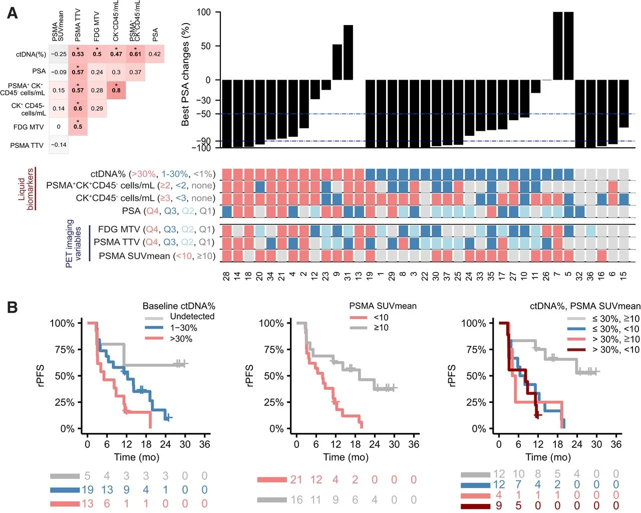
Among the baseline parameters evaluated, ctDNA fraction and PSMA SUVmean were the only variables associated with rPFS, while baseline ctDNA was also associated with overall survival.
Patients with ctDNA <30% and PSMA SUVmean ≥10 experienced the longest rPFS, with the median not reached, compared with approximately 5–8 months among patients with ctDNA ≥30%, PSMA SUVmean <10, or both adverse features. Because of the small cohort, the study was not sufficiently powered to demonstrate independent prognostic value through multivariable analysis, but the pattern suggests that these measurements provide complementary information.
Their biological meanings are different. ctDNA fraction provides a circulating measure of tumor-derived molecular burden, while PSMA PET measures expression of the therapeutic target across metastatic lesions.
The relationship between them was also notable. Median ctDNA was 26.9% in patients with PSMA SUVmean <10 versus 4.2% in those with PSMA SUVmean ≥10. Thus, high circulating tumor burden could coexist with relatively low PSMA avidity, potentially identifying a disease state in which tumor burden is increasing while the target required for efficient radioligand delivery is less strongly expressed.
TP53, RB1 and PTEN Alterations Are Linked to a Metabolically Aggressive Phenotype
Genomic profiling further separated tumors with different biological behavior.
Alterations involving TP53, RB1 and PTEN were particularly enriched among patients with poor outcomes. Seven of eight ctDNA-evaluable patients with primary resistance, defined as rPFS <3 months, harbored alterations in these tumor-suppressor genes.
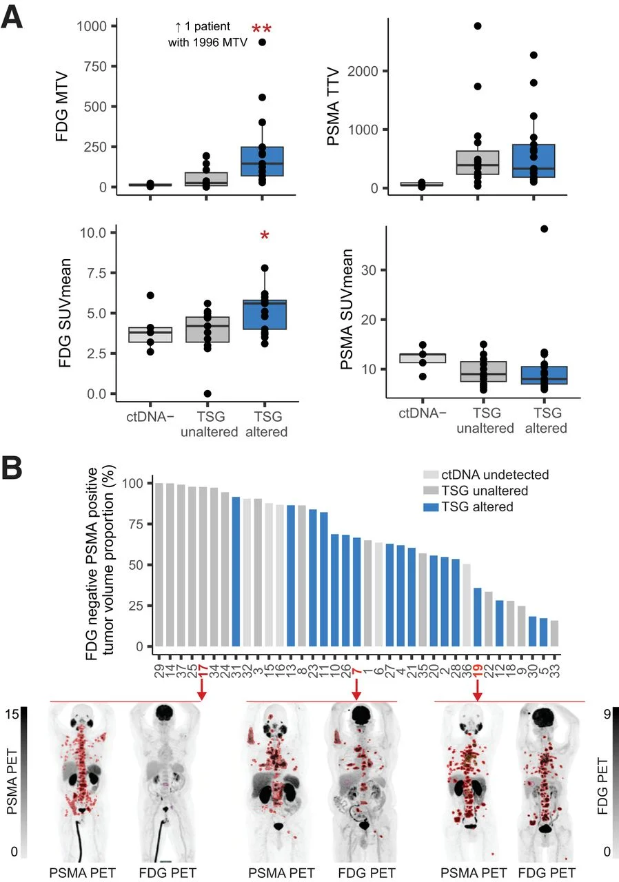
These genomic alterations were associated with a distinct metabolic phenotype on ^18F-FDG PET. Patients with tumor-suppressor gene alterations had a median ^18F-FDG metabolic tumor volume of 145 mL versus 30 mL in those without such alterations (P=0.004), and median ^18F-FDG SUVmean was 5.6 versus 4.2 (P=0.03). By contrast, PSMA tumor volume and PSMA SUVmean were similar between the two groups.
This dissociation is biologically important. Comparable PSMA expression does not necessarily imply comparable tumor behavior. Two tumors may present a similar amount of target for ^177Lu-PSMA-617 while differing substantially in genomic aggressiveness and metabolic activity.
The study therefore positions FDG PET as more than another measure of tumor volume. In this cohort, metabolic activity was linked to an aggressive genomic state characterized by tumor-suppressor loss.
There were also potentially favorable genomic observations. ATM and SPOP alterations occurred exclusively among patients whose rPFS exceeded the cohort median, although the numbers were too small for definitive conclusions. One patient with biallelic MSH2 loss and high TMB experienced a >99% PSA reduction and PFS exceeding 12 months despite high baseline ctDNA and low PSMA SUVmean. These findings are hypothesis-generating and cannot establish predictive effects of these individual alterations.
ctDNA at Week 12 Provides an Early Molecular Readout of Treatment Efficacy
The longitudinal analysis produced one of the clearest signals in the study.
At 12 weeks, ctDNA fell below the study’s detection threshold of 1% in 17 of 35 patients (49%). Among patients with undetectable ctDNA, 94% achieved PSA90, compared with only 6% of patients in whom ctDNA remained detectable.
Persistent ctDNA was also strongly associated with shorter rPFS, with an HR of 6.3 (95% CI 2.5–15.7; P<0.001).
PSMA PET response assessed using RECIP classification was independently informative. PSA90 rates were 83% among patients with partial response, 41% among those with stable disease and 0% among those with progressive disease. RECIP progression was also associated with markedly shorter rPFS.
The most interesting observation emerged when these two measurements were evaluated together.
Discordance Between ctDNA and PSMA PET May Reveal Residual Resistant Disease
Imaging response and molecular response were not always equivalent.
Among patients classified as having stable disease by PSMA PET RECIP, ctDNA identified strikingly different response states. PSA90 occurred in 100% of patients with undetectable ctDNA compared with only 9% of those with persistent ctDNA.
Two illustrative patients demonstrated more than a 30% reduction in PSMA-positive total tumor volume at week 12 but retained detectable ctDNA. Both subsequently experienced short rPFS.
This discordance has important implications for monitoring PSMA-directed treatment. A reduction in PSMA-positive disease on imaging may accurately demonstrate elimination of treatment-sensitive lesions while failing to capture molecular persistence of resistant tumor populations.
ctDNA, conversely, does not provide spatial information or directly measure PSMA expression, but it can integrate tumor-derived genomic material originating from multiple metastatic sites.
The two approaches therefore interrogate different components of response. PSMA PET measures what is happening to target-expressing disease; ctDNA may indicate whether molecular disease persists despite that imaging response.
Resistance Involves Both Phenotypic PSMA Loss and Genomic Clonal Selection
Serial analysis at progression revealed that resistance was accompanied by changes in both target expression and tumor genomic composition.
Most patients had lower PSMA SUVmean and SUVmax at progression than at baseline. The reduction was particularly pronounced among patients whose tumors had high PSMA avidity before treatment. This pattern is compatible with treatment-mediated selection or expansion of PSMA-low tumor populations.
Genomic evolution occurred in parallel.
Among 25 patients with detectable ctDNA at both baseline and progression, most clonal mutations remained stable, whereas mutations that changed substantially in cancer-cell fraction were more frequently subclonal at baseline. Importantly, 5 of 25 patients (20%) demonstrated clonal expansion of nonsynonymous tumor-suppressor alterations, involving TP53 in three patients, RB1 in one and PTEN in one.
The same alterations associated with aggressive disease at baseline could therefore become increasingly represented within the tumor population during treatment.
This provides a model of resistance in which therapy does not simply fail uniformly across the tumor. Instead, treatment applies selective pressure to a heterogeneous cancer population, reducing sensitive clones while allowing pre-existing or emerging resistant populations to expand.
PSMA Loss Appears Predominantly Phenotypic Rather Than Genomic
One of the most intriguing observations concerns the mechanism underlying declining PSMA expression.
A patient who developed a FOLH1 alteration at progression also showed reduced PSMA PET uptake and a shift from PSMA-positive to PSMA-negative circulating prostate cancer cells. FOLH1 encodes PSMA, making this an informative example of genomic evolution directly involving the therapeutic target.
However, this was not the dominant pattern.
Across most patients, reduced PSMA expression at progression occurred without corresponding genomic alterations in FOLH1. The authors therefore suggest that PSMA expression may be predominantly regulated through nongenetic mechanisms, potentially including epigenetic regulation.
Single-cell CTC analysis provided additional support for this interpretation. PSMA-positive and PSMA-negative circulating prostate cancer cells did not demonstrate consistent genomic differences, indicating that PSMA phenotype cannot simply be inferred from the underlying genomic clone.
This distinction may become increasingly important as PSMA-directed therapies move into broader treatment settings. Target expression itself is dynamic, and resistance may develop through phenotypic adaptation without requiring permanent loss or alteration of the gene encoding the target.
What Do These Biomarkers Actually Predict?
For an immuno-oncology interpretation, this is the critical unresolved question.
The study demonstrates that low baseline ctDNA, high PSMA avidity, early ctDNA clearance and PSMA PET response are associated with favorable outcomes during ^177Lu-PSMA-617 plus pembrolizumab. It does not, however, establish that these are biomarkers of sensitivity to the combination specifically.
Every patient in PRINCE received both treatments. There was no ^177Lu-PSMA-617-alone control group and no external cohort treated with the same combination. Consequently, the contribution of pembrolizumab cannot be separated from the activity of the radiopharmaceutical.
This limitation is particularly important because the translational analysis focused predominantly on tumor-intrinsic biomarkers. It provides extensive information on ctDNA, CTC genomics, PSMA expression and metabolic phenotype, but limited insight into T-cell activation, immune-cell composition or other host immune features that might specifically explain the contribution of PD-1 blockade.
The ongoing EVOLUTION trial, randomizing patients with advanced mCRPC to ^177Lu-PSMA-617 versus ^177Lu-PSMA-617 plus ipilimumab/nivolumab, is expected to incorporate both tumor-intrinsic and immune correlatives. Such a randomized framework should provide a better opportunity to determine which biomarkers reflect general radioligand sensitivity and which identify biological interaction with immune checkpoint inhibition.
You Can Read Full Article Here