
PD-L1 expression has become one of the most widely used biomarkers in cancer immunotherapy, but its clinical interpretation is more complicated than simply classifying a tumor as positive or negative. In non-small-cell lung cancer (NSCLC), treatment selection often relies on the Tumor Proportion Score (TPS), while in head and neck, gastric, and several other malignancies, the Combined Positive Score (CPS) plays an important role. In other settings, particularly historical atezolizumab trials in triple-negative breast cancer (TNBC), PD-L1 expression has been assessed using an Immune Cell (IC) score.
This raises an important question: why does the same biomarker require different scoring systems across cancer types, and why can a tumor be considered PD-L1-positive for one immunotherapy but not another?
The explanation lies in the interaction between tumor biology, immune-cell distribution, diagnostic technology, and clinical evidence. PD-L1 is expressed not only by malignant cells but also by immune populations within the tumor microenvironment, and these cellular compartments may contribute differently to immune regulation. However, the clinical adoption of TPS, CPS, and IC has been shaped not only by biological considerations but also by the assays and thresholds used in pivotal immunotherapy trials.
Understanding these differences is essential because TPS, CPS, and IC measure different aspects of PD-L1 expression, and their numerical values are not interchangeable.

Lung Cancer Hope Month 2026: Celebrating Progress in Modern Oncology
Why Do Tumors Express PD-L1?
The PD-1/PD-L1 pathway is a physiological mechanism that helps regulate immune activation and prevent excessive tissue damage. PD-1 is an inhibitory receptor expressed by activated T cells, while PD-L1 can be expressed by malignant cells, macrophages, dendritic cells, and other populations within the tumor microenvironment.
When PD-L1 binds PD-1, inhibitory signaling suppresses pathways involved in T-cell receptor activation and CD28 costimulation, reducing T-cell proliferation, cytokine production, and cytotoxic activity. Tumors can exploit this mechanism to weaken immune responses directed against malignant cells, allowing cancer cells to survive despite the presence of immune recognition.
An important scientific distinction is that PD-L1 expression may arise through different biological processes. Some tumors express PD-L1 through intrinsic oncogenic signaling, including pathways involving PI3K–AKT or other molecular alterations, whereas others increase PD-L1 expression in response to inflammatory signals generated by an ongoing immune response.
The latter mechanism, known as adaptive immune resistance, was described in influential research by Taube and colleagues, published in Science Translational Medicine in 2012. The investigators demonstrated that PD-L1 expression could be spatially associated with immune infiltrates, supporting the concept that tumors may upregulate inhibitory signaling in response to local immune activation.
Mechanistically, activated tumor-reactive T cells release interferon-γ, which stimulates PD-L1 expression through the JAK–STAT pathway, particularly STAT1 and IRF1. This creates a biological feedback loop in which T cells recognize malignant cells and generate inflammation, but the resulting PD-L1 expression can suppress further T-cell activity.
Consequently, PD-L1 positivity does not always indicate the same immune environment. In one tumor, it may reflect an active but inhibited antitumor response, while in another, PD-L1 expression may occur without substantial infiltration by functional tumor-reactive lymphocytes. This distinction helps explain why PD-L1 expression alone cannot fully predict response to checkpoint inhibition and why assessing different cellular compartments may provide different clinical information.
What Is TPS, and What Does It Actually Measure?
The Tumor Proportion Score measures the percentage of viable tumor cells demonstrating partial or complete membranous PD-L1 staining at any intensity. It is calculated by dividing the number of PD-L1-positive tumor cells by the total number of viable tumor cells and multiplying by 100. Importantly, PD-L1 expression on lymphocytes, macrophages, and other immune populations is excluded from this calculation.
For example, a tumor specimen containing 100 viable cancer cells, of which 60 demonstrate PD-L1 membrane staining, would have a TPS of 60%, regardless of how many surrounding immune cells express PD-L1. TPS therefore provides a direct measurement of PD-L1 expression by malignant cells, although it cannot determine whether that expression is driven by oncogenic signaling or induced by an ongoing immune response.
The clinical importance of TPS became particularly evident in NSCLC, where tumor-cell PD-L1 expression was prospectively evaluated during the development of pembrolizumab. In the phase III KEYNOTE-024 trial, published by Reck and colleagues in the New England Journal of Medicine in 2016, patients with previously untreated advanced NSCLC, PD-L1 TPS ≥50%, and no sensitizing EGFR mutations or ALK rearrangements were randomized to pembrolizumab or platinum-based chemotherapy. Pembrolizumab significantly improved median progression-free survival from 6.0 to 10.3 months and demonstrated an overall survival benefit, establishing high tumor-cell PD-L1 expression as an important treatment-selection biomarker.
The subsequent KEYNOTE-042 trial extended the investigation to patients with TPS ≥1%, although the magnitude of treatment benefit differed across PD-L1 expression subgroups. These findings reinforced the importance of interpreting PD-L1 not simply as positive or negative but in relation to specific expression thresholds and treatment strategies.
The reason TPS became established in NSCLC is therefore closely connected to clinical validation. Tumor-cell PD-L1 expression was the scoring approach used in pivotal pembrolizumab trials, and it successfully identified populations in which treatment provided meaningful benefit. This does not mean that immune-cell PD-L1 is biologically irrelevant in lung cancer. Macrophages, dendritic cells, and lymphocytes contribute substantially to immune regulation, but their PD-L1 expression is not incorporated into TPS.
Importantly, not all immunotherapy-containing NSCLC regimens require PD-L1 positivity. Several chemotherapy–immunotherapy combinations have demonstrated benefit across PD-L1 expression groups, illustrating that the predictive importance of TPS depends on whether immunotherapy is administered alone or alongside other treatments.
What Is CPS, and Why Does It Include Immune Cells?
The Combined Positive Score evaluates PD-L1 expression in both malignant cells and selected tumor-associated immune populations. It is calculated by adding the number of PD-L1-positive tumor cells, lymphocytes, and macrophages, dividing this sum by the total number of viable tumor cells, and multiplying by 100. The reported score is capped at 100, even when the number of positive tumor and immune cells exceeds the number of viable tumor cells.
Unlike TPS, CPS incorporates PD-L1 expression in the immune compartment, which may be particularly informative in tumors where immune cells contribute substantially to local inhibitory signaling.
For example, a tumor specimen containing 100 viable cancer cells, of which only two express PD-L1, may also contain 18 PD-L1-positive lymphocytes and macrophages. Although the TPS would be just 2%, the CPS would reach 20 because immune-cell PD-L1 expression contributes to the numerator. This illustrates how the same tumor can demonstrate low PD-L1 expression among malignant cells but a higher combined score when the surrounding immune environment is considered.
Biologically, CPS may capture features of adaptive immune resistance that are not fully represented by tumor-cell staining alone. Activated T cells can stimulate PD-L1 expression on nearby immune populations through interferon-γ signaling, creating an inhibitory environment that limits further T-cell activity. However, CPS does not directly measure immune activation, nor does a high score necessarily indicate the presence of functional tumor-reactive lymphocytes.
Its clinical relevance has emerged most clearly in head and neck, gastric, gastroesophageal, cervical, and selected breast cancer settings, where pivotal trials evaluated PD-L1 expression using combined tumor and immune-cell scoring.
Why Is CPS Important in Head and Neck Cancer?
Head and neck squamous cell carcinoma (HNSCC) frequently develops within a complex immune microenvironment influenced by chronic inflammation, tumor-associated macrophages, lymphocyte infiltration, and, in some cases, HPV-associated antigens. PD-L1 expression may occur in malignant cells as well as immune populations surrounding tumor nests, making combined assessment biologically plausible.
The clinical importance of CPS was demonstrated in the phase III KEYNOTE-048 trial, published by Burtness and colleagues in The Lancet in 2019. The study compared pembrolizumab alone, pembrolizumab plus chemotherapy, and cetuximab plus chemotherapy in recurrent or metastatic HNSCC.
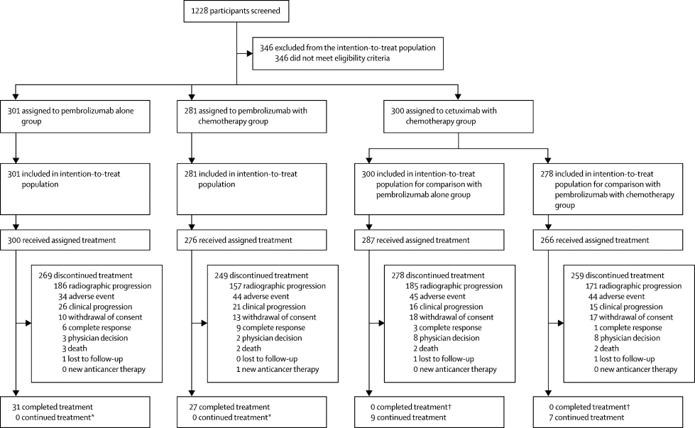
Pembrolizumab monotherapy significantly improved overall survival among patients with CPS ≥20, with median survival of 14.9 months compared with 10.7 months for cetuximab plus chemotherapy. A survival benefit was also demonstrated in patients with CPS ≥1, with median overall survival of 12.3 versus 10.3 months.
These findings established CPS as an important biomarker for pembrolizumab monotherapy selection. However, the trial also demonstrated that pembrolizumab plus chemotherapy improved overall survival in the broader study population, illustrating that the importance of PD-L1 selection differs according to the treatment regimen.
From an immunological perspective, CPS may capture PD-L1 expression associated with both tumor cells and infiltrating immune populations, but KEYNOTE-048 did not prove that immune-cell PD-L1 independently determines sensitivity to pembrolizumab. Its clinical adoption reflects the evidence generated using the combined scoring method rather than proof that CPS is universally superior to TPS in head and neck cancer.
Why Is CPS Used in Gastric and Gastroesophageal Cancer?
Gastric and gastroesophageal cancers are characterized by substantial biological heterogeneity, including differences in molecular subtype, immune infiltration, stromal composition, and inflammatory signaling. PD-L1 expression may be concentrated in immune cells surrounding malignant glands rather than exclusively within tumor cells, making CPS a potentially informative approach to assessing the tumor–immune environment.
The phase III CheckMate 649 trial, published in The Lancet in 2021, provided important evidence supporting the clinical relevance of CPS in advanced gastric, gastroesophageal junction, and esophageal adenocarcinoma. The trial evaluated nivolumab plus chemotherapy versus chemotherapy alone and demonstrated improved overall survival and progression-free survival in patients with PD-L1 CPS ≥5.
In the primary analysis, median overall survival was 14.4 months with nivolumab plus chemotherapy compared with 11.1 months with chemotherapy alone. The benefit remained evident with longer follow-up, including the five-year analysis published in 2026, which reported five-year overall survival rates of 16% versus 6% in the CPS ≥5 population.
These results highlight how CPS can identify clinically relevant populations in upper gastrointestinal cancer. Nevertheless, the threshold is not universal, and the predictive importance of CPS may differ across checkpoint inhibitors and treatment combinations. Trials such as KEYNOTE-859 have further demonstrated that the magnitude of immunotherapy benefit can vary according to PD-L1 expression, emphasizing the need to interpret each scoring threshold within the context of the regimen and clinical evidence supporting it.
The use of CPS in gastric cancer therefore reflects both biological considerations and prospective trial validation. Although immune-cell PD-L1 expression may contribute to the immunological environment, there is no established rule that CPS is intrinsically more biologically accurate than TPS for every gastric tumor.
What Is the IC Score, and Why Is It Different From CPS?
The Immune Cell score focuses on PD-L1 expression within tumor-associated immune populations rather than malignant cells. In the VENTANA SP142 scoring algorithm, which became particularly important in trials of atezolizumab, IC is defined by the percentage of the assessed tumor area occupied by PD-L1-positive immune cells.
This is fundamentally different from TPS and CPS because IC is an area-based measurement rather than a cell-count ratio. While TPS evaluates the proportion of viable tumor cells expressing PD-L1 and CPS incorporates selected PD-L1-positive immune cells into a combined calculation, the SP142 IC algorithm estimates the extent of PD-L1-positive immune-cell staining within the tumor area.
Consequently, the hypothetical specimen described earlier, with a TPS of 2% and a CPS of 20, cannot be assigned an IC score from cell counts alone. Its IC classification would depend on the area occupied by the PD-L1-positive immune cells, including their distribution within the assessed tumor tissue.
This distinction is particularly important because a tumor with relatively few PD-L1-positive immune cells distributed over a large area may be classified differently from one containing densely clustered positive immune cells. IC scoring therefore provides another perspective on immune-cell-associated PD-L1 expression, although it does not directly measure immune-cell function or the strength of antitumor immunity.
Why Did IC Scoring Become Important in Triple-Negative Breast Cancer?
Triple-negative breast cancer provides one of the most informative examples of how different PD-L1 scoring systems can emerge within the same malignancy.
TNBC is often associated with higher levels of tumor-infiltrating lymphocytes than hormone receptor-positive breast cancer, although immune infiltration varies considerably between tumors. PD-L1 expression can be prominent in tumor-associated immune cells, including macrophages and lymphocytes, creating a biological rationale for evaluating the immune compartment.
The phase III IMpassion130 trial, published by Schmid and colleagues in the New England Journal of Medicine in 2018, investigated atezolizumab plus nab-paclitaxel in previously untreated unresectable locally advanced or metastatic TNBC. PD-L1 positivity was defined using the VENTANA SP142 assay, with a threshold of IC ≥1%, meaning that PD-L1-positive immune cells occupied at least 1% of the assessed tumor area.
The trial demonstrated improved progression-free survival with atezolizumab plus nab-paclitaxel, including among patients classified as PD-L1-positive. These findings supported the accelerated US approval of the combination in 2019, making IC scoring an important part of the historical development of immunotherapy in TNBC.
However, the atezolizumab indication for metastatic TNBC was voluntarily withdrawn in the United States in 2021. IMpassion130 therefore remains a landmark biomarker-development study rather than an example of a currently approved US treatment indication.
The most interesting scientific question is whether IC scoring was selected because PD-L1 expression on immune cells was biologically more important for atezolizumab activity than expression on malignant cells. Although immune-cell PD-L1 is relevant to local inhibitory signaling, the trial did not establish that IC scoring was inherently superior to CPS or that atezolizumab specifically required immune-cell PD-L1 expression.
Instead, the SP142 assay and its IC algorithm were part of the clinical development program, and the resulting biomarker-defined population was evaluated prospectively alongside treatment outcomes.
Why Did KEYNOTE-355 Use CPS Instead of IC in the Same Cancer?
The difference becomes particularly interesting when IMpassion130 is compared with the phase III KEYNOTE-355 trial, which evaluated pembrolizumab plus chemotherapy in previously untreated locally recurrent inoperable or metastatic TNBC.
Unlike IMpassion130, KEYNOTE-355 assessed PD-L1 expression using the 22C3 assay and CPS, incorporating PD-L1-positive tumor cells and selected immune cells into the same calculation.
The overall survival analysis, published in the New England Journal of Medicine in 2022, demonstrated that pembrolizumab plus chemotherapy significantly improved survival in patients with CPS ≥10. Median overall survival was 23.0 months compared with 16.1 months for chemotherapy alone, corresponding to a hazard ratio of 0.73.
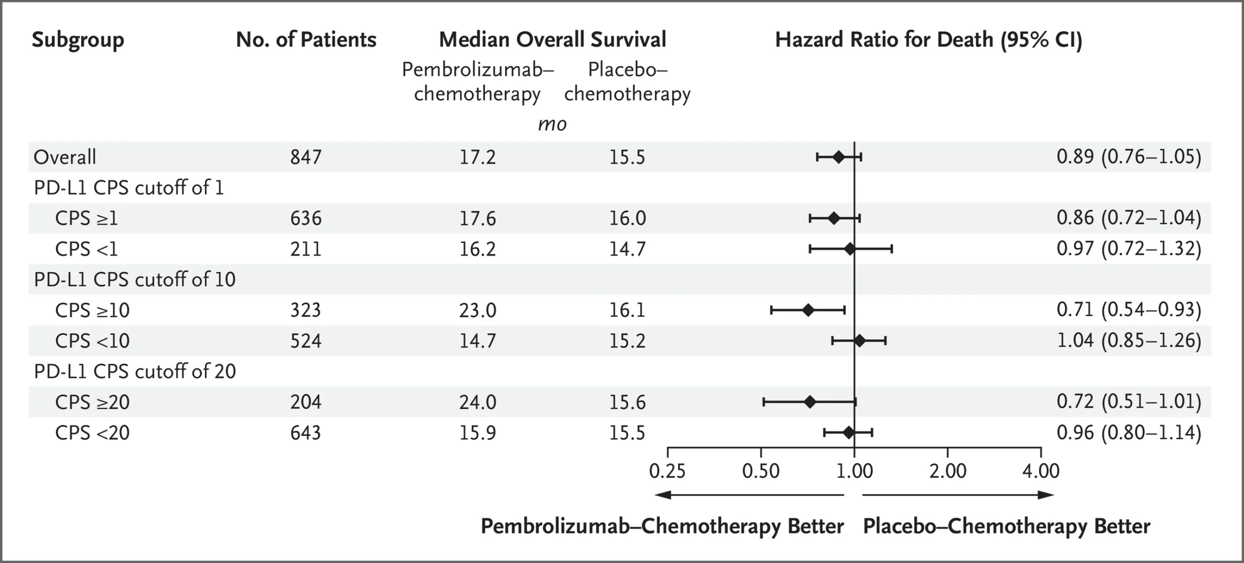
Interestingly, the broader CPS ≥1 population did not demonstrate a statistically significant overall survival improvement, with median survival of 17.6 versus 16.0 months.
These findings illustrate that the threshold used to define PD-L1 positivity can influence the identification of patients who derive clinically meaningful benefit. A lower cutoff includes more patients, but it may also include tumors in which the relationship between PD-L1 expression and treatment benefit is weaker.
The comparison between IMpassion130 and KEYNOTE-355 raises a broader question about biomarker development. Both trials investigated checkpoint inhibition in metastatic TNBC, yet one used SP142 IC ≥1% while the other used 22C3 CPS ≥10. This difference should not be interpreted as evidence that atezolizumab biologically requires immune-cell PD-L1 while pembrolizumab requires combined tumor and immune-cell PD-L1.
The two trials evaluated different checkpoint inhibitors, chemotherapy backbones, assays, and scoring algorithms, and they were not designed to establish which PD-L1 measurement was universally superior. Nevertheless, their results demonstrate why PD-L1 positivity is not a single interchangeable classification, even within the same tumor type.
Why Can the Same Cancer Use Different PD-L1 Scores?
The experience in TNBC demonstrates that the choice of PD-L1 scoring system is influenced by more than cancer histology. Different antibody clones and staining platforms can produce different PD-L1 detection patterns, while the scoring algorithm determines whether expression is assessed in malignant cells, immune cells, or both. The clinical significance of the resulting measurement is then evaluated in trials using particular checkpoint inhibitors, treatment combinations, and PD-L1 thresholds.
This means that the same tumor may meet the criteria for PD-L1 positivity in one clinical development program but not another, without either result necessarily being incorrect. The assays may simply be measuring different cellular compartments or applying different thresholds to the observed staining.
An important example comes from urothelial carcinoma, where checkpoint inhibitor development programs historically employed different PD-L1 assays and scoring methods. Atezolizumab trials used SP142-based immune-cell assessment, while pembrolizumab studies used CPS-based testing. Durvalumab development also involved tumor-cell and immune-cell PD-L1 assessment.
These approaches reflected the diagnostic strategies used in individual clinical trials rather than a universal biological rule that urothelial cancer should be assessed by one specific score.
The practical implication is that a CPS of 10 in TNBC cannot automatically be considered equivalent to a CPS of 10 in gastric cancer, and neither result can be translated into a TPS of 10% in NSCLC. Even when numerical values appear similar, the underlying measurements and their validated clinical meanings may differ.
Does the PD-L1 Antibody Clone Matter?
An additional challenge is that PD-L1 immunohistochemistry is not performed using a single standardized antibody or staining platform. Commonly used assays include 22C3, 28-8, SP263, and SP142, each developed with particular diagnostic procedures and clinical applications.
The Blueprint PD-L1 IHC Assay Comparison Project, published by Hirsch and colleagues in the Journal of Thoracic Oncology in 2017, provided important evidence about the comparability of these assays in NSCLC.
The investigators found that 22C3, 28-8, and SP263 generally demonstrated similar tumor-cell staining patterns, whereas SP142 tended to identify fewer PD-L1-positive tumor cells. Greater variability was observed in immune-cell staining and its interpretation, indicating that agreement between assays was less consistent when the immune compartment was assessed.
These findings are clinically important because a tumor classified as PD-L1-positive using one assay may not receive the same classification with another, particularly when expression is close to a treatment-selection threshold. Differences in staining sensitivity, the cellular compartment assessed, and observer interpretation can influence the final result.
However, analytical agreement between assays does not automatically establish clinical interchangeability. Even when two assays produce similar staining results, the clinical performance of one cannot be assumed to match the other unless appropriate validation has been performed.
This is why PD-L1 testing should be interpreted in relation to the diagnostic assay and scoring algorithm associated with the treatment indication, rather than treating all PD-L1 measurements as equivalent.
Why Do Some PD-L1-Negative Tumors Still Respond to Immunotherapy?
Despite its widespread clinical use, PD-L1 remains an imperfect predictive biomarker. Some patients with high PD-L1 expression do not respond to checkpoint inhibitors, while others with low or undetectable expression may still experience meaningful and durable benefit.
One explanation is that PD-L1 expression does not directly measure whether functional tumor-reactive T cells are present. A tumor may express PD-L1 through oncogenic signaling but lack the immune infiltration necessary for effective PD-1 blockade. Conversely, a tumor with relatively low PD-L1 expression may contain immunogenic neoantigens and activated immune populations capable of responding when inhibitory signaling is interrupted.
This distinction was explored in influential research by Tumeh and colleagues, published in Nature in 2014. The investigators studied patients with melanoma receiving pembrolizumab and found that clinical responses were associated with pre-existing CD8-positive T cells located at the invasive tumor margin, together with evidence of an adaptive immune response.
The study helped establish an important concept: successful PD-1 blockade may depend not simply on the presence of PD-L1 but on the existence of a tumor-reactive immune response that can be reinvigorated.
Tumor heterogeneity introduces further complexity because PD-L1 expression may vary between different regions of the same lesion, between primary and metastatic sites, and over time following systemic therapy or radiation. A small biopsy may therefore capture only part of the tumor’s immune environment, and a result obtained before treatment may not fully represent the biology of disease at progression.
Additional mechanisms can also limit checkpoint inhibitor activity despite high PD-L1 expression. Defective antigen presentation, loss of interferon signaling, T-cell exclusion, immunosuppressive myeloid populations, and alternative inhibitory pathways may prevent effective antitumor responses even when PD-1/PD-L1 signaling is blocked.
PD-L1 expression should therefore be understood as one component of a broader immunological picture rather than a complete measure of checkpoint inhibitor sensitivity.
Could Spatial Biology Improve PD-L1 Assessment?
One of the most interesting developments in immuno-oncology is the growing recognition that the location of PD-L1 expression may be as important as its overall abundance.
Conventional TPS, CPS, and IC scoring provide clinically useful measurements, but they cannot fully describe how PD-L1-positive tumor cells, macrophages, dendritic cells, and lymphocytes are organized within the tumor microenvironment. Two tumors with similar PD-L1 scores may have very different immune architectures, including differences in T-cell proximity to malignant cells, macrophage distribution, and the presence of immunosuppressive stromal regions.
Spatial transcriptomics and multiplex immunohistochemistry offer opportunities to investigate these relationships by preserving tissue architecture while identifying cellular populations and molecular signaling patterns.
For example, PD-L1 expression concentrated at the interface between tumor cells and activated T cells may reflect adaptive immune resistance, whereas diffuse expression in a tumor with limited lymphocyte infiltration may represent a different biological state. Similarly, PD-L1-positive macrophages positioned close to dysfunctional T cells may contribute to inhibitory signaling that cannot be fully characterized by a single numerical score.
Recent studies using spatial profiling have demonstrated that the organization of immune cells can be associated with response to checkpoint inhibition, supporting the idea that immune-cell location and interaction may provide information beyond conventional biomarker expression.
However, these technologies remain investigational for routine treatment selection, and prospective validation is required before spatial immune signatures can replace or supplement established PD-L1 diagnostic algorithms.
The future of PD-L1 assessment may therefore involve integrating conventional scoring with immune-cell phenotypes, tumor antigenicity, functional T-cell states, and spatial relationships rather than relying on a single measurement.