
The central challenge of cancer screening is simple to describe but extraordinarily difficult to solve: find the tumor while it is still small enough to cure.
Most emerging blood-based approaches attempt to detect something released directly by cancer, including circulating tumor DNA, abnormal methylation patterns, proteins or other tumor-derived molecules. But early cancers create a fundamental technical problem. A tiny tumor may shed only minute quantities of material into the circulation, leaving the diagnostic signal buried beneath an enormous background of normal DNA and proteins.
The immune system approaches the problem differently.
A developing cancer does not necessarily need to release enough material into blood for us to measure it directly. It only needs to produce enough abnormal biology for the immune system to notice. Tumor-associated or mutation-derived antigens can be presented to T cells, specific T-cell clones can expand, B cells can generate antibodies, and systemic immune populations can begin to change. Each responding lymphocyte can then proliferate, potentially amplifying a biological signal that originated from a very small number of malignant cells.
This creates a provocative possibility: the immune system may sometimes detect cancer before our conventional technologies can detect the tumor itself.
If so, early cancer detection could eventually involve more than searching blood for fragments of cancer. We might also search for evidence that the immune system has already encountered it.
The Immune System as a Biological Amplifier
The difference between tumor-derived biomarkers and immune-derived biomarkers is fundamental.
Consider circulating tumor DNA. A malignant cell must release DNA into the circulation, that DNA must survive degradation, enter the blood sample and then be captured by an assay sensitive enough to distinguish it from an overwhelming background of normal cell-free DNA. When tumors are very small, this becomes increasingly difficult simply because there may be too few tumor-derived molecules available.
Adaptive immunity can amplify weak biological signals.
A dendritic cell presenting a tumor-associated peptide can activate a rare antigen-specific T cell. That cell can proliferate into thousands or millions of descendants sharing related antigen specificity. B cells can similarly undergo clonal expansion and produce large quantities of antibodies against tumor-associated antigens.
The original malignant signal may therefore be tiny, while the immune response generated against it can be much larger.
This is the central rationale behind immune-based early detection. Rather than asking only whether we can detect the cancer, we ask whether we can detect the host’s response to cancer.
The concept is not entirely new. Tumor-associated autoantibodies have been investigated for decades. What has changed is our ability to interrogate immune responses at enormous scale through T-cell receptor sequencing, B-cell repertoire analysis, proteomics, seromics and machine learning.
The immune fingerprint of an early cancer may no longer need to be a single biomarker. It can be a pattern.
Every T Cell Carries a Record of What It Has Recognized
T cells recognize peptide antigens through the T-cell receptor, or TCR. Because TCRs are generated through somatic recombination, the human T-cell repertoire contains an enormous diversity of receptor sequences.
When a T cell encounters its cognate antigen, that clone can expand. The composition of the circulating TCR repertoire therefore contains information about immune encounters occurring throughout the body.
In principle, cancer should leave traces in this repertoire.
The challenge is that the signal is extraordinarily complex. A blood sample contains T cells responding to previous infections, vaccination, chronic viruses, autoantigens, environmental exposures and many other stimuli. Cancer-specific clones represent only a fraction of this immunological history.
Recent computational approaches are beginning to extract the relevant patterns.
In 2024, researchers reported iCanTCR in Cancer Research, a deep-learning framework trained using more than 2,000 TCR repertoires spanning 11 cancer types and healthy controls. The model used peripheral-blood TCRβ sequences to estimate cancer probability and achieved an area under the receiver operating characteristic curve of 0.86 for identifying early-stage cancers in the evaluated datasets.
The importance of the study was conceptual as much as technical. It suggested that the circulating T-cell repertoire contains sufficient information for an algorithm to distinguish at least some patients with early cancer from individuals without cancer.
But an important question remained: are these algorithms recognizing genuine tumor-directed immune biology, or simply finding indirect differences between patient populations?
More recent studies have begun to address that problem.
In 2025, TCR Sequencing Began to Look Much More Biological
A particularly important study appeared in npj Precision Oncology in July 2025: “Circulating T-cell Receptor Repertoire for Cancer Early Detection,” by Yilong Li, Michelle Nahas, Roman Yelensky and colleagues.
The investigators studied 463 patients with lung cancer, 86% of whom had stage I disease, together with 587 individuals without known cancer. Instead of looking only for individual TCR sequences, they organized related receptors into what they called TCR repertoire functional units, or RFUs, based on sequence similarity.
The rationale was biologically attractive. Different TCR sequences can potentially recognize the same or related antigens. Grouping receptors according to sequence relationships may therefore capture immune responses that would be missed if every TCR were treated as an isolated event.
The investigators identified 327 cancer-associated RFUs. Importantly, 247 of those 327 RFUs were associated with specific HLA alleles, supporting the idea that antigen presentation was contributing to the observed TCR patterns. Tumor-infiltrating lymphocyte TCRs from several of the identified groups also bound HLA-presented tumor antigen peptides.
This moves the observation beyond a purely statistical fingerprint. At least part of the circulating TCR signal appears to reflect genuine antigen recognition.
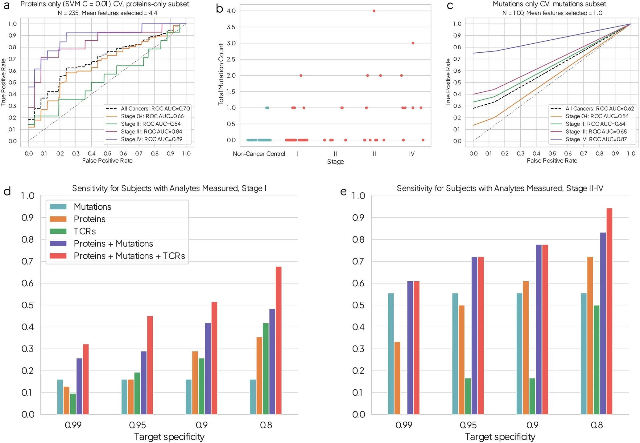
The resulting immune score detected approximately 48% of stage I lung cancers at 80% specificity. That performance alone would not be sufficient for a population-wide screening test, particularly because screening demands extremely careful control of false positives. But another result was more interesting: when the TCR score was combined with circulating tumor DNA and protein biomarkers, sensitivity increased by as much as 20 percentage points in the evaluated multi-analyte setting.
That may point toward the future of early detection.
The immune system may not replace tumor-derived biomarkers. It may detect something different from them.
Tumor DNA and Immune Memory Are Looking at Different Sides of the Same Cancer
A ctDNA assay asks whether malignant cells are shedding detectable molecular material into the bloodstream.
A TCR assay asks whether the adaptive immune system has recognized something sufficiently abnormal to alter its repertoire.
These signals are related, but they are not equivalent.
A small tumor might release very little DNA yet generate an immunogenic antigen capable of expanding specific T-cell populations. Conversely, a tumor could shed measurable DNA while remaining relatively poorly immunogenic.
Combining tumor-derived and host-response signals therefore makes biological sense. One measures the cancer itself; the other measures the organism’s reaction to it.
This distinction became particularly interesting in 2025 when researchers demonstrated, using prospectively collected samples from the Atherosclerosis Risk in Communities cohort, that mutations detectable by a multicancer early-detection assay near the time of diagnosis could in some individuals be traced back in plasma more than three years before clinical cancer diagnosis, although at dramatically lower allele fractions.
The study, published in Cancer Discovery, involved small numbers and was designed to establish biological feasibility rather than screening performance. Nevertheless, it illustrates the central technical challenge of detecting cancer directly at its earliest stages: the signal is already present, but it can be extraordinarily faint.
The immune system offers another way to approach that faint signal.
Instead of measuring how much tumor DNA exists, we may be able to measure whether the host has amplified recognition of the abnormal cells.
Nasopharyngeal Cancer Offers a Remarkable Natural Experiment
One of the most interesting demonstrations came from nasopharyngeal carcinoma, where Epstein-Barr virus creates an unusual opportunity to study immune recognition before clinical diagnosis.
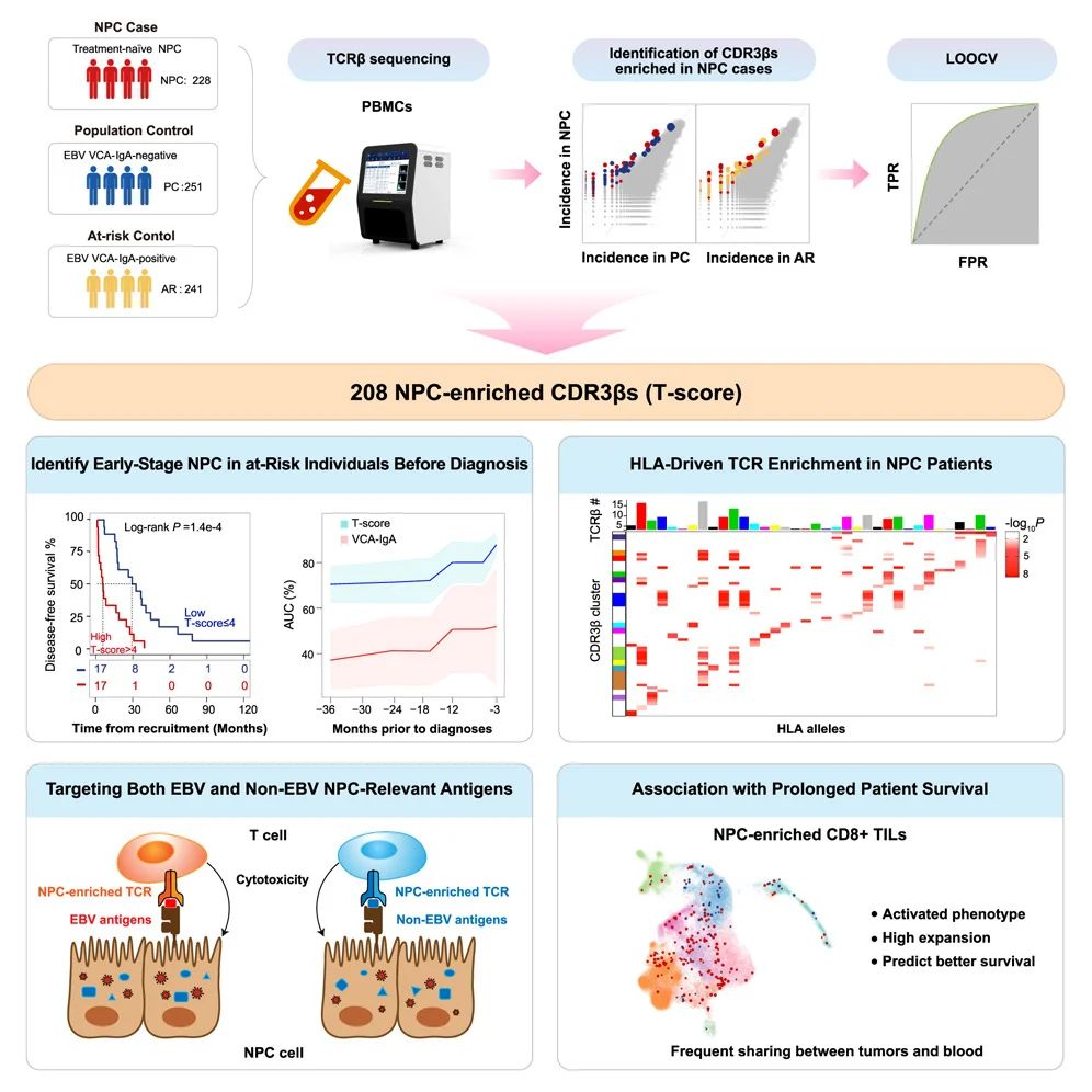
In 2025, investigators reported “Immunosequencing Identifies Signatures of T Cell Responses for Early Detection of Nasopharyngeal Carcinoma” in Cancer Cell.
They identified 208 TCRβ CDR3 sequences enriched in nasopharyngeal carcinoma and used them to construct a TCR-based T-score. The signature distinguished patients with cancer and, importantly, identified early-stage disease among EBV-seropositive individuals at elevated risk.
The biological analysis was particularly compelling. The cancer-associated TCRs were not directed exclusively against EBV. They also recognized nonviral antigens expressed by the malignant cells, suggesting that the circulating immune repertoire was capturing a broader antitumor response rather than simply measuring chronic viral immunity.
Higher T-scores were associated with shorter intervals to eventual cancer diagnosis in at-risk individuals, providing evidence that the immune signal evolved as clinically detectable cancer approached.
This is exactly the kind of observation that makes immune-based screening interesting. The blood was not simply revealing that EBV existed. It was revealing that the immune system was responding differently as cancer developed.
The Immune Fingerprint May Appear Before the Tumor Is Diagnosed
The strongest evidence for the concept comes from longitudinal samples collected before cancer diagnosis.
Cross-sectional studies can show that patients with stage I cancer differ immunologically from healthy controls, but that does not prove the immune alteration preceded clinical disease. Differences could reflect the presence of an already detectable tumor, associated inflammation or other factors.
Prediagnostic samples provide a much harder test.
A 2026 study in the British Journal of Cancer examined circulating tumor-associated autoantibodies in small-cell lung cancer and lung adenocarcinoma. Researchers analyzed serum from 695 individuals and identified distinct autoantibody panels for the two lung cancer subtypes.
The most intriguing part of the study involved serial samples collected before diagnosis.
In patients with available longitudinal sera, tumor-associated autoantibody concentrations increased as diagnosis approached. Several individuals had detectable responses more than two years before clinical diagnosis, and overall 67% of the autoantibody-positive patients evaluated longitudinally were already panel-positive at least six months before diagnosis.
The numbers in the longitudinal analysis were small, so these findings require larger prospective validation. They nevertheless demonstrate an important biological principle: humoral recognition of tumor-associated antigens can precede the moment at which cancer becomes clinically diagnosed.
Cancer may therefore leave an immunological trail before it leaves an obvious clinical one.
Antibodies Have One Major Advantage: Amplification
Autoantibodies are particularly attractive for early detection because B cells provide enormous biological amplification.
A tumor-associated protein may exist at concentrations too low to detect reliably in plasma. But if the immune system recognizes that protein as abnormal, activated B cells can generate large quantities of antibodies against it. Those antibodies can remain stable in circulation even when the antigen itself is scarce.
The immune system has effectively performed part of the amplification step before the blood sample reaches the laboratory.
This phenomenon has been observed previously in several cancers. TP53 autoantibodies, for example, have been detected before conventional biomarkers in subsets of patients with ovarian cancer, while autoantibody responses against cancer-associated proteins have been identified years before diagnosis in longitudinal breast and gastric cancer cohorts.
The 2026 lung cancer study adds contemporary molecular resolution to this older concept. The investigators found that autoantibody induction was related not only to antigen abundance but also to somatic alterations and HLA class II variation, again suggesting that the signal reflects an interaction between tumor biology and the patient’s immune system rather than a generic inflammatory response.
That interaction may ultimately prove more informative than any individual autoantibody.
The Fingerprint May Be Personal Because HLA Is Personal
One challenge in immune-based cancer detection is also one of its most interesting features.
Different people do not necessarily respond to the same tumor antigen in the same way.
T-cell recognition depends on antigen presentation by HLA molecules, and HLA genes are among the most polymorphic loci in the human genome. A tumor peptide efficiently presented by one person’s HLA molecules may be poorly presented in another individual.
The 2025 npj Precision Oncology TCR study illustrates this clearly: approximately three quarters of the cancer-associated TCR functional units identified were associated with particular HLA alleles.
This means that a universal cancer-associated TCR signature may be difficult to construct. But it also provides biological information. If an algorithm knows a person’s HLA genotype, it may be able to interpret the repertoire in the context of which tumor antigens that immune system is capable of presenting.
Future immune-screening algorithms might therefore not ask simply whether a particular TCR is present.
They could ask: given this person’s HLA genotype, age, immune history and TCR repertoire, is the pattern of clonal expansion consistent with recognition of an emerging malignancy?
That is a much more personalized form of screening.
Can the Immune Repertoire Help Us Decide Whether a Lung Nodule Is Cancer?
Early detection does not always mean finding an invisible tumor. Sometimes the clinical problem is determining whether something already visible on imaging is malignant.
Low-dose CT screening can detect very small pulmonary nodules, but many are benign. This creates a different diagnostic challenge: how do we identify which nodules require invasive investigation or treatment?
TCR profiling is now being explored here as well.
A large 2025 Cancer Research study performed TCR profiling using 6,059 blood samples and 988 tumor samples and developed the LungTCR database. The investigators then constructed TCRnodseek plus, integrating TCR features with clinical information and CT imaging.
The model was evaluated prospectively across multiple centers in 1,107 patients with indeterminate pulmonary nodules, providing a substantially larger validation setting than many earlier immune-repertoire studies.
The significance of this approach is not simply that another algorithm classified nodules. It demonstrates how immune information could complement radiology. CT describes the physical lesion. The TCR repertoire potentially describes whether the host immune system is reacting to biology associated with malignancy.
The combination of what the lesion looks like and how the immune system responds to it may ultimately be more informative than either alone.
Breast Cancer May Also Be Visible in the Circulating TCR Repertoire
The concept is not restricted to lung cancer.
A 2025 study in npj Systems Biology and Applications examined peripheral blood TCR repertoires in women with and without breast cancer. Machine-learning analysis distinguished cancer status with an average AUC of approximately 0.96 in the study population.
The cohort was small, containing 98 women, and therefore cannot establish screening performance in the general population. But the study adds another independent observation: tumor-associated information can be extracted from circulating TCR repertoires across different cancer types.
This raises a much larger question.
If different cancers generate partially distinct immune responses, could one blood sample eventually identify not only whether cancer may be present, but also which tissue is generating the immune signal?
That is considerably harder.
T cells respond to antigens, not anatomical labels. Shared tumor-associated antigens, viral exposures, HLA diversity and cross-reactivity can all blur tissue specificity. Determining the likely tissue of origin may therefore require integration of immune information with tumor-derived signals such as methylation, fragmentomics or circulating proteins.
The immune fingerprint may be best suited to answering one part of a multi-layered diagnostic question.
AI Is Becoming Necessary Because the Fingerprint Is Too Complex to Read Manually
A single TCR sequence tells us very little without knowing its antigen specificity. A human repertoire can contain millions of distinct clonotypes, many at extremely low abundance.
This is why machine learning has become central to immune-based detection.
iCanTCR used deep learning to identify cancer-associated repertoire patterns. DeepCaTCR, reported in 2025, combined convolutional neural networks, bidirectional long short-term memory and attention mechanisms to recognize cancer-associated TCR features. In its reported datasets, the approach achieved high specificity and an AUC of 0.967 for its pan-cancer repertoire score, although sensitivity for early-stage disease remained substantially lower and independent prospective validation will be essential.
These algorithms are not discovering a hidden conventional biomarker. They are attempting to recognize distributed patterns across thousands or millions of immune receptors.
The distinction from our AI target-discovery question is important. Here, AI is not being used to identify a new immunotherapy target. It is functioning more like a translator, attempting to convert the immune system’s response history into a clinically interpretable signal.
The biological information already exists.
The challenge is learning how to read it.
But Infection, Aging and Autoimmunity Also Leave Immune Fingerprints
This is where the concept becomes difficult.
Cancer is not the only process that changes the immune repertoire.
Viral and bacterial infections produce enormous T-cell expansions. Vaccination alters immune clonotypes. Autoimmune diseases generate antigen-specific responses. Aging changes repertoire diversity and clonal hematopoiesis can alter circulating immune populations. Smoking, chronic inflammatory diseases, immunosuppressive medications and previous cancers can all affect immune composition.
A screening test must therefore distinguish cancer-associated immune recognition from the enormous background of normal and pathological immune history.
This is much harder than distinguishing a group of known cancer patients from young healthy controls.
The clinically relevant populations are older adults with pulmonary disease, infections, autoimmune disorders, previous malignancies and multiple comorbidities. These are exactly the individuals in whom false-positive immune signals could become problematic.
The 2025 lung cancer TCR study attempted to address some of this complexity by including individuals undergoing low-dose CT screening or bronchial evaluation rather than relying entirely on idealized healthy controls. The ability of its TCR score to distinguish lung cancer from benign pulmonary nodules was therefore particularly relevant.
Still, prospective population-level validation will be essential before immune-repertoire tests can be considered screening tools.
Screening Creates a Statistical Problem Biology Cannot Solve Alone
Even an impressive diagnostic AUC does not automatically translate into a useful cancer-screening test.
Cancer prevalence in an asymptomatic screening population is generally low. When prevalence is low, even a relatively small false-positive rate can generate many more false positives than true cancers.
Specificity therefore becomes critical.
Consider a hypothetical test applied to 100,000 asymptomatic individuals in whom 1% actually have the cancer being screened for. At 90% sensitivity and 95% specificity, the test would identify most cancers, but it would also produce thousands of false-positive results.
Those individuals may then undergo imaging, repeated blood tests, biopsies or invasive procedures.
This is why early-stage case-control performance should never be equated automatically with validated screening utility.
For immune-based detection, the evidence currently supports biological and translational promise, not replacement of established screening programs.
The field still needs prospective studies in intended-use populations showing that immune signatures can improve clinically meaningful detection without generating unacceptable false-positive rates.
Perhaps We Should Not Ask the Immune System to Do Everything
The most compelling future may therefore be multi-analyte detection.
Tumor DNA provides direct molecular evidence of malignancy. Methylation patterns can provide information about tissue of origin. Fragmentomics can detect changes in the physical characteristics of circulating DNA. Proteins capture altered tumor and host biology. Imaging provides anatomical information.
Immune profiling adds another dimension: evidence that the host has recognized abnormal biology.
The 2025 circulating TCR study already provides a proof of principle for this complementarity. Adding its TCR score to ctDNA and circulating protein biomarkers increased sensitivity for early-stage lung cancer in the evaluated subset.
This may be particularly valuable when tumor shedding is low. An early cancer could produce insufficient ctDNA for confident detection but enough antigenic stimulation to generate a measurable adaptive immune response.
Conversely, an immune-silent tumor might be detected through DNA or protein signals even when no strong immune fingerprint exists.
Rather than searching for one perfect biomarker, early detection may therefore become a process of combining independent biological views of the same developing cancer.
Could the Immune System Detect Precancer?
An even more provocative question lies one step earlier.
If immune recognition can occur before invasive cancer becomes clinically apparent, could immune profiling detect the transition from normal tissue to premalignancy?
In principle, immune surveillance begins as abnormal cells acquire mutations, altered protein expression, genomic instability or viral oncogenic programs. Some of these changes could generate antigens before a lesion has developed the biological characteristics required for invasive cancer.
But this creates an interpretive problem.
Not every abnormal clone becomes cancer. Many premalignant lesions remain stable or regress. Ageing tissues accumulate mutations without inevitably progressing to malignancy. Detecting an immune response to abnormal cells could therefore identify biological risk without identifying a cancer that actually requires treatment.
The most useful immune fingerprint may consequently be one that does more than detect immune recognition. It would need to distinguish productive immune surveillance of a lesion that will never progress from an evolving immune response to a lesion becoming clinically dangerous.
Longitudinal studies will be essential to answer this question.
A static blood sample may tell us that the immune system has noticed something.
Repeated measurements may tell us whether that something is changing.
The Trajectory May Matter More Than the Snapshot
The 2026 autoantibody study offers an important clue. In prediagnostic lung cancer samples, antibody levels did not simply appear as a binary positive or negative signal. They tended to increase as clinical diagnosis approached.
The nasopharyngeal cancer TCR study similarly linked stronger TCR-based signals with shorter intervals to eventual diagnosis.
This suggests that immune trajectories may contain more information than single measurements.
A stable immune signature over five years could represent previous infection, benign inflammation or controlled premalignancy. A rapidly expanding set of tumor-associated TCR clonotypes or rising autoantibody response could indicate an evolving antigenic process.
Screening might therefore eventually resemble monitoring rather than one-time classification.
Instead of asking: “Is cancer present today?”
an immune-based test might ask: “Has this person’s immune response changed in a way that suggests an emerging malignancy?”
That is a fundamentally different approach to early detection.
Could Screening Become Personalized to an Individual’s Immune Baseline?
Current screening tests generally compare an individual with population-derived thresholds. Immune profiling creates the possibility of comparing someone partly with their own previous immune state.
Every person has a different repertoire shaped by HLA genotype, age, infections, vaccination, environmental exposure and genetics. This heterogeneity makes population-based immune biomarkers difficult.
But longitudinal measurement could turn heterogeneity into an advantage.
If a baseline repertoire were available, new clonal expansions, changing TCR functional units, emerging autoantibodies or shifts in immune-cell states could be identified relative to that person’s previous profile.
The clinically relevant signal might therefore not be: “Your immune system looks like that of people with cancer.”
It could eventually become: “Your immune system is beginning to recognize something it was not recognizing before.”
Whether such longitudinal immune surveillance will be technically feasible, cost-effective or sufficiently specific remains unknown. But conceptually it may fit the biology of early cancer better than forcing every individual into a universal immune threshold.
What Would It Take to Turn an Immune Fingerprint Into a Screening Test?
The gap between an interesting biomarker and a useful screening test is large.
First, immune signatures must be validated prospectively in the populations in which they would actually be used. Studies need appropriate controls with infections, inflammatory disorders, benign lesions and other cancers.
Second, models must remain robust across age, ancestry, HLA genotype and environmental exposures. This is particularly important for TCR-based approaches because HLA diversity directly influences antigen presentation and repertoire structure.
Third, the test must provide clinically actionable information. A positive immune signal without a way to localize the cancer could create uncertainty and unnecessary diagnostic procedures.
Fourth, investigators must establish the lead time gained by immune detection. Finding a cancer two weeks before imaging would have little clinical value. Detecting biologically meaningful disease months or years earlier could potentially transform outcomes, but only if intervention at that earlier stage improves health rather than producing overdiagnosis.
Finally, screening trials must evaluate outcomes rather than AUC alone. The ultimate question is not whether an algorithm can separate stored blood samples from cancer cases and controls. It is whether using the test in asymptomatic people can find consequential cancers earlier while keeping harms acceptable.
That standard has not yet been met for immune-repertoire screening.