






Every day, millions of patients undergo blood tests. But what if that same blood sample could tell us much more than we imagine?
Among billions of blood cells, extremely rare tumor cells may be hiding. Circulating Tumor Cells (CTCs) can be very few, yet extraordinarily rich in information. CTCs are tumor cells that detach from the primary tumor or metastatic sites and enter the bloodstream. They represent a minimally invasive source of tumor-derived information and a unique window into tumor biology. As key players in metastasis, CTCs can provide valuable insights into tumor burden, metastatic dissemination, treatment response, and tumor heterogeneity.
Over the past two decades, CTCs have emerged as an important component of liquid biopsy. Unlike circulating tumor DNA (ctDNA) and many other circulating biomarkers, CTCs are complete tumor cells carrying the full biological complexity of the cancer, providing access not only to its genetic alterations but also to its molecular, phenotypic, and morphological characteristics.
This allows them to be counted, characterized at the morphological and molecular levels, cultured in the laboratory, and even used to investigate drug sensitivity. In other words, CTCs do not simply tell us how much tumor is present. They can provide clues about what the tumor looks like, how it is changing, and how it might behave. And we can get all of this information from something as simple and familiar as a routine blood draw. This combination of a minimally invasive sampling method and the wealth of biological information that CTCs can provide is what makes them one of the most intriguing tools in liquid biopsy.
But What Do We Know About the Clinical Significance of CTCs
The clinical relevance of CTCs has been extensively investigated across several solid tumors, with the strongest and most consistent evidence coming from metastatic breast, prostate, and colorectal cancer. The CellSearch® system, the only FDA-approved liquid biopsy test (Ref.1), is one of the most extensively studied methods. This technology exploits the expression of epithelial markers, particularly epithelial cell adhesion molecules (EpCAM), to enrich tumor cells, followed by their identification using markers such as cytokeratins and CD45. In these settings, CTC enumeration has repeatedly demonstrated prognostic value: patients with higher baseline CTC counts generally have shorter progression-free and overall survival, while changes in CTC levels during treatment can provide additional information about disease course and prognosis.
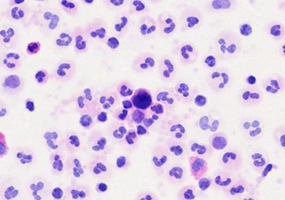
Picture courtesy of Tethis S.p.A.
In metastatic breast cancer, CTC counts have been extensively studied and are consistently associated with both progression-free and overall survival (Ref.1). Similar findings have been reported in metastatic castration-resistant prostate cancer (Ref.2), where CTC numbers and changes during treatment have been shown to provide important prognostic information and have been investigated as markers of treatment response and disease progression. In metastatic colorectal cancer, CTC levels have also been associated with survival outcomes, although their integration into routine clinical management remains more limited (Ref.3).
But How Close are We to Bringing the Potential of CTCs Into Clinical Practice?
A biomarker may have strong prognostic value without necessarily being useful for selecting the most effective treatment. In other words, knowing that a patient has a poorer prognosis does not necessarily tell us which treatment is most likely to work for that individual. Predicting the course of the disease is not the same as guiding treatment decisions.
The SWOG S0500 trial is a good example. In patients with metastatic breast cancer who continued to have high CTC counts after starting first-line chemotherapy, changing treatment early based on the CTC result did not improve overall survival compared with continuing the same treatment until conventional signs of disease progression appeared. CTCs could identify patients with a poorer prognosis, but using this information alone to change treatment did not make a difference in how patients ultimately did. (Ref.4).
More recent studies suggest that the picture may be more nuanced. The STIC CTC trial in metastatic breast cancer showed that CTC counts could potentially help guide the choice between endocrine therapy and chemotherapy in selected patients. This suggests that CTCs may have clinical value when they are used to answer a specific treatment question, rather than simply predicting prognosis. (Ref 5-6).
But turning this potential into a useful clinical tool means overcoming several biological, technical, and clinical challenges.
One of the most important challenges is the very low abundance of CTCs in peripheral blood. In many patients with solid tumors, CTCs may occur at extremely low frequencies among millions of leukocytes, making their reliable detection technically demanding. Consequently, the analytical sensitivity of the assay can substantially influence the reported CTC count. A low or undetectable CTC count does not necessarily indicate the biological absence of tumor cells in the circulation, but may instead reflect limitations in blood volume analyzed, enrichment efficiency, or the biological characteristics of the tumor cells. This issue becomes particularly relevant in early-stage disease and in tumor types characterized by low CTC shedding.
A second major limitation is the marked biological heterogeneity of CTCs. CTCs are not a homogeneous population: they may differ in epithelial and mesenchymal phenotype, expression of tumor-associated markers, viability, genomic alterations, and metastatic potential. In particular, epithelial–mesenchymal transition (EMT) may result in reduced expression of epithelial markers such as EpCAM, which are commonly exploited for CTC enrichment. Therefore, EpCAM-dependent approaches may preferentially capture epithelial CTCs while underrepresenting mesenchymal or hybrid CTC populations. Conversely, label-free approaches based on physical properties such as size or deformability may recover different CTC subpopulations and may introduce their own selection biases. As a consequence, different technologies can measure partially different biological compartments rather than the same universal CTC population. (Ref.7)
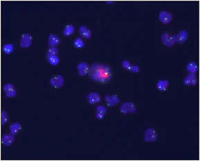
Picture courtesy of Tethis S.p.A.
A further challenge arises from the interaction between CTC biology and detection technology. Since CTCs constitute a highly heterogeneous population, no single method is able to capture all clinically relevant CTC subsets with equal efficiency. Different assays therefore use different enrichment strategies and identification criteria, each introducing its own selection bias. Consequently, CTC platforms may measure partially different biological populations, making cross-study comparisons difficult and hindering methodological standardization.
So, Where Do We Stand Today?
The challenge is no longer simply to show that CTCs can be detected or that they are associated with patient outcomes. The real question is whether, and in which situations, CTC information can actually change treatment decisions and improve patient outcomes. The future of CTCs in the clinic will likely depend on moving beyond a simple cell count and towards a more comprehensive, multiparametric view of the circulating tumor cell population, integrating CTCs with other components of liquid biopsy.
And this brings us back to the fundamental challenge: billions of cells travel through the bloodstream, yet only a small fraction may carry critical information about the disease. Finding these rare cells requires methods that are sufficiently sensitive to detect them and sufficiently sophisticated to distinguish and characterize those that truly matter.
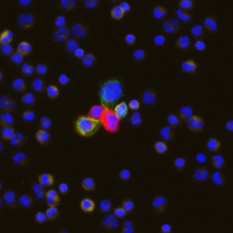
Picture courtesy of Tethis S.p.A.
Getting there will require more standardized and sensitive methods, better characterization of clinically relevant CTC populations, and prospective clinical trials designed to determine whether CTC-guided decisions can actually improve patient outcomes. But characterization does not have to stop at counting or molecular profiling. We can also look at the cells themselves: their size, shape, nuclear features, and other cytological characteristics.
Because if every cell can tell a different story, why just count CTCs when we can look them in the face?
References
- Cristofanilli M, et al. Circulating tumor cells, disease progression, and survival in metastatic breast cancer. N Engl J Med. 2004;351:781–791.
- de Bono JS, et al. Circulating tumor cells predict survival benefit from treatment in metastatic castration-resistant prostate cancer. Clin Cancer Res. 2008;14:6302–6309.
- Cohen SJ, et al. Relationship of circulating tumor cells to tumor response, progression-free survival, and overall survival in patients with metastatic colorectal cancer. J Clin Oncol. 2008.
- Smerage JB, et al. Circulating Tumor Cell Measurement to Assess Tumor Response to Therapy and Predict Survival in Metastatic Breast Cancer. J Clin Oncol. 2014;32:3483–3489.
- Bidard FC, et al. Efficacy of Circulating Tumor Cell Count-Driven vs Clinician-Driven First-line Therapy Choice in Hormone Receptor-Positive, ERBB2-Negative Metastatic Breast Cancer: The STIC CTC Randomized Clinical Trial. JAMA Oncol. 2021;7:34–41.
- Bidard FC, et al. Overall Survival With Circulating Tumor Cell Count-Driven Choice of Therapy in Advanced Breast Cancer: A Randomized Trial. J Clin Oncol. 2024.
- Rushton AJ, et al. Review of Circulating Tumour Cell Enrichment Technologies. Cancers. 2021.
The article is supported by Tethis S.p.A.