Combining tumor-derived extracellular vesicles with circulating tumor cells improved prognostic stratification in patients with metastatic breast cancer, according to an analysis of two independent patient cohorts.
The study identified more than 20 tumor-derived extracellular vesicles per 7.5 mL of blood as a clinically relevant threshold associated with poorer survival. Importantly, elevated tumor-derived extracellular vesicles identified higher-risk patients even when conventional circulating tumor cell counts were low.
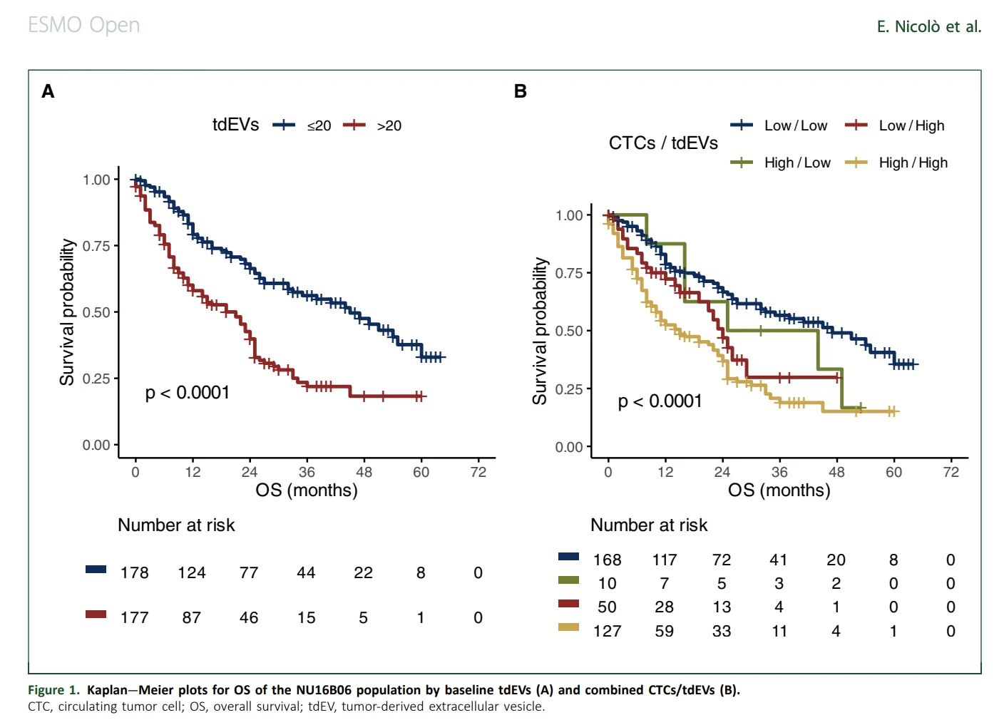
In the training cohort, median overall survival was 21 months among patients with elevated tumor-derived extracellular vesicles and 45 months among those with lower levels. Patients with high levels of both biomarkers had the shortest median overall survival at 15 months, compared with 47 months among patients with low levels of both.
The findings, published in ESMO Open, support tumor-derived extracellular vesicles as complementary prognostic biomarkers rather than replacements for circulating tumor cell enumeration. Prospective studies are still needed before the combined assay can guide routine treatment decisions (Nicolò et al., 2026).
Why Are Additional Biomarkers Needed in Metastatic Breast Cancer?
Treatment selection in metastatic breast cancer has become increasingly complex as endocrine therapies, molecularly targeted agents, antibody–drug conjugates, immunotherapies, and HER2-directed treatments move across different disease settings.
Tumour subtype remains fundamental, but patients with similar clinicopathological characteristics can experience substantially different disease courses. Blood-based biomarkers could provide additional information about disease burden and biological aggressiveness without requiring repeated tissue biopsies.
Circulating tumor cells are among the most extensively studied liquid-biopsy biomarkers in metastatic breast cancer. Using the CellSearch system, a threshold of five circulating tumor cells per 7.5 mL of blood has been validated for prognostic stratification.
However, some patients with fewer than five circulating tumor cells still experience aggressive disease. Nicolò and colleagues investigated whether tumor-derived extracellular vesicles could reveal prognostic information not captured by circulating tumor cells alone (Nicolò et al., 2026).
What Are Tumor-Derived Extracellular Vesicles?
Extracellular vesicles are membrane-bound particles released by cells into the surrounding environment. Tumor-associated extracellular vesicles can carry proteins, lipids, nucleic acids, and other biological material involved in intercellular communication.
In cancer, these particles have been linked to tumour growth, metastatic spread, modification of the tumour microenvironment, and treatment resistance.
The particles evaluated in this study were large extracellular vesicle-like objects isolated alongside circulating tumor cells after epithelial cell adhesion molecule enrichment. They were identified through CellSearch images using the open-source Automated CTC Classification, Enumeration and PhenoTyping software, known as ACCEPT.
The investigators defined circulating tumor cells as EpCAM-positive, cytokeratin-positive, DAPI-positive, and CD45-negative particles larger than 4 μm. Tumor-derived extracellular vesicles were classified as EpCAM-positive, cytokeratin-positive, DAPI-negative, and CD45-negative particles smaller than 14 μm.
The authors emphasized that these particles were identified through imaging, phenotypic, and morphometric criteria. They were not comprehensively molecularly characterized according to current International Society for Extracellular Vesicles recommendations and are therefore more accurately regarded as operationally defined tumour-associated extracellular vesicle-like particles (Nicolò et al., 2026).
How Was the Study Conducted?
The retrospective observational analysis included two independent cohorts.
The training cohort consisted of 355 patients with metastatic breast cancer enrolled in the NU16B06 study at Northwestern University between 2016 and 2021. Patients could have any breast cancer subtype and were included if blood had been collected before beginning a new treatment line and archived CellSearch images were available.
Approximately 53% had hormone receptor-positive, HER2-negative disease, 19% had HER2-positive breast cancer, and 27% had triple-negative breast cancer. Forty-three percent had at least three metastatic sites, while 67% had visceral metastases.
Most patients were beginning first- or second-line treatment. Therapies included chemotherapy, endocrine therapy with or without targeted agents, HER2-directed treatment, and immunotherapy-containing regimens.
The predefined tumor-derived extracellular vesicle threshold was then evaluated in an independent group of 385 patients from the phase 3 STIC CTC trial. Unlike the training cohort, this validation population consisted of patients with hormone receptor-positive, HER2-negative metastatic breast cancer receiving first-line treatment (Nicolò et al., 2026).
Why Was a Threshold of 20 Extracellular Vesicles Selected?
The investigators examined several possible thresholds, including the median value, percentile-based divisions, previously published categories, and an analytically derived optimal value of 67 tumor-derived extracellular vesicles.
Higher levels were associated with shorter overall survival across multiple thresholds. The statistical performance of the different models was also similar, with Brier Score differences below 0.005.
The researchers selected a single threshold of more than 20 tumor-derived extracellular vesicles per 7.5 mL of blood because earlier studies had identified 0–20 as a reference range among healthy donors. The threshold was also considered easier to integrate into a two-biomarker model with the established circulating tumor cell cutoff.
Patients were therefore categorized as having low tumor-derived extracellular vesicle levels at 20 or fewer particles and high levels at more than 20 particles per 7.5 mL (Nicolò et al., 2026).
What Did Extracellular Vesicle Levels Show in the Training Cohort?
The median tumor-derived extracellular vesicle count in the NU16B06 cohort was 20 per 7.5 mL of blood.
Patients with more than 20 particles had a median overall survival of 21 months, compared with 45 months among patients with 20 or fewer.
The univariable hazard ratio for death was 2.38, indicating more than twice the mortality risk in the elevated group. After adjustment for circulating tumor cells, breast cancer subtype, treatment line, metastatic distribution, and other clinical factors, elevated tumor-derived extracellular vesicles remained independently associated with poorer survival:
- Adjusted HR 1.65; 95% CI, 1.09–2.52; p=0.019
Circulating tumor cell elevation also remained independently prognostic, although its adjusted effect was slightly smaller:
- Adjusted HR 1.51; 95% CI, 1.02–2.25; p=0.040
These results indicate that the two biomarkers captured overlapping but not identical prognostic information (Nicolò et al., 2026).
What Happened When Both Biomarkers Were Combined?
Combining the two measurements divided patients into four prognostic groups.
Patients with fewer than five circulating tumor cells and no more than 20 tumor-derived extracellular vesicles had the most favorable outcome, with a median overall survival of 47 months.
Patients with at least five circulating tumor cells and more than 20 tumor-derived extracellular vesicles had the poorest outcome, with a median overall survival of 15 months.
In the multivariable model, the group with high levels of both biomarkers had a 2.49-fold higher risk of death than the low-circulating tumor cell, low-extracellular vesicle group:
- HR 2.49; 95% CI, 1.74–3.56
The survival curves presented on page 4 of the publication show the clearest separation for the group with simultaneous elevation of circulating tumor cells and tumor-derived extracellular vesicles.
Could Extracellular Vesicles Identify Risk When CTC Counts Were Low?
This was one of the most clinically relevant findings.
Among 218 patients with fewer than five circulating tumor cells, 23% had elevated tumor-derived extracellular vesicle levels. In the unadjusted analysis, these patients had a higher risk of death than those with low levels of both biomarkers:
- HR 1.78; 95% CI, 1.09–2.88; p=0.020
After adjustment for clinical and pathological variables, the association weakened and was no longer statistically significant:
- HR 1.53; 95% CI, 0.93–2.53
The result suggests that extracellular vesicles can reveal heterogeneity within a group conventionally considered to have a more favorable prognosis. However, the loss of statistical significance in the adjusted training-cohort analysis means the finding required independent validation.
Were the Results Confirmed in the STIC CTC Cohort?
The threshold was successfully validated in the independent STIC CTC cohort.
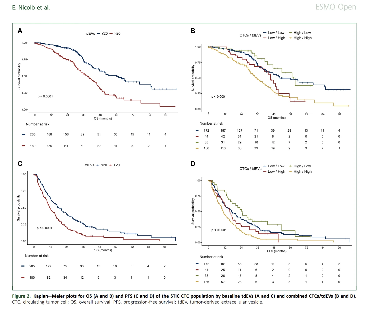
Patients with more than 20 tumor-derived extracellular vesicles had significantly poorer overall survival. The association remained statistically significant after adjustment:
- HR 2.28; 95% CI, 1.52–3.34; p<0.001
Among patients with low circulating tumor cell counts, elevated tumor-derived extracellular vesicles were associated with a nearly twofold increase in mortality risk:
- HR 1.96; 95% CI, 1.15–3.33
Patients with high levels of both biomarkers again had the poorest outcome:
- HR 2.74; 95% CI, 1.82–4.11
By contrast, patients with high circulating tumor cell counts but low extracellular vesicle levels did not have significantly poorer overall survival than patients with both biomarkers low:
- HR 0.96; 95% CI, 0.50–1.84
The Kaplan–Meier plots on page 7 show consistent separation for both overall survival and progression-free survival according to extracellular vesicle status. The high-circulating tumor cell, high-extracellular vesicle group had the least favorable outcomes, while elevated extracellular vesicles continued to identify poorer prognosis among patients with low circulating tumor cell counts (Nicolò et al., 2026).
Were Extracellular Vesicles Detected Without Circulating Tumor Cells?
Tumor-derived extracellular vesicles were more frequently detectable than circulating tumor cells.
In the NU16B06 cohort, 87 patients, representing 25% of the population, had no detectable circulating tumor cells. Tumor-derived extracellular vesicles were still identified in this group, with a median count of two particles per 7.5 mL.
Only 31 patients, or 9%, had no detectable tumor-derived extracellular vesicles.
Counts of the two biomarkers were strongly correlated, with a Spearman coefficient of 0.796, but the ability to detect extracellular vesicles in patients without circulating tumor cells supports their potential complementary role.
The broader numerical range of extracellular vesicles could also provide more material for future analyses of tumour-associated markers, although this application was not directly validated in the study.
Did Breast Cancer Subtype Influence Extracellular Vesicle Levels?
Higher tumor-derived extracellular vesicle counts were observed in invasive lobular carcinoma than in ductal carcinoma.
The investigators noted that higher circulating tumor cell counts have previously been described in invasive lobular breast cancer, potentially because reduced cell-to-cell adhesion facilitates the release of tumour material into the circulation.
Tumor-derived extracellular vesicle levels were also lower in HER2-positive disease than in hormone receptor-positive, HER2-negative and triple-negative breast cancer within the heterogeneous training cohort.
These findings were exploratory. The study was not designed to establish subtype-specific thresholds, and the biological mechanism underlying the higher extracellular vesicle levels in invasive lobular carcinoma remains uncertain.
Can the Biomarker Combination Guide Treatment?
The study demonstrates prognostic value, but it does not establish predictive value.
A prognostic biomarker provides information about expected outcomes. A predictive biomarker identifies whether a patient is more likely to benefit from a particular treatment.
Patients were not randomly assigned to treatment according to tumor-derived extracellular vesicle levels. The analysis therefore cannot show that patients with both biomarkers elevated would benefit from treatment escalation or that patients with both biomarkers low could safely receive less intensive therapy.
The authors proposed that the combined assay could eventually support risk-adapted clinical trial designs. Patients with low levels of both biomarkers could potentially be studied in treatment-de-escalation strategies, while those with high levels could be candidates for treatment-intensification trials.
These applications remain hypotheses requiring prospective clinical validation.
What Are the Advantages of the Combined Approach?
Both biomarkers were obtained from the same 7.5-mL blood sample and analyzed within the CellSearch imaging framework.
Automated ACCEPT analysis showed strong agreement with manually counted circulating tumor cells. The correlation between automated and manual circulating tumor cell counts was 0.848, and both approaches produced similar survival associations.
Automation could reduce operator variability and improve reproducibility when processing large numbers of samples. It could also increase the amount of information extracted from existing CellSearch images without requiring a separate blood collection.
However, technical standardization, regulatory validation, laboratory availability, and cost-effectiveness must be assessed before routine implementation.
What Are the Main Limitations?
The analysis was retrospective, even though blood collection in the contributing studies was prospective. Only a subset of the STIC CTC trial population had the required images and complete data, creating a possibility of selection bias.
The two cohorts were also clinically different. The training population included all metastatic breast cancer subtypes and multiple treatment lines, whereas the validation cohort included only hormone receptor-positive, HER2-negative disease treated in the first-line setting before CDK4/6 inhibitors became standard.
The results might therefore not apply equally to every contemporary metastatic breast cancer population.
The study evaluated baseline samples only. It did not determine whether changes in tumor-derived extracellular vesicles during treatment could predict response or progression.
Circulating tumour DNA was not available, preventing direct comparison or integration with one of the most widely used contemporary liquid-biopsy biomarkers.
The extracellular vesicle-like particles were defined by CellSearch and ACCEPT imaging characteristics rather than comprehensive molecular confirmation of their tumour origin. The selected threshold requires further prospective and analytical validation before it can be considered a standardized clinical test.
Funding and Disclosures
The STIC CTC trial was funded by Institut Curie, the French National Cancer Institute, and Menarini Silicon Biosystems. Eleonora Nicolò received support from an American-Italian Cancer Foundation postdoctoral research fellowship.
Several authors disclosed research relationships, consulting roles, speaker fees, or other professional relationships with pharmaceutical, diagnostic, and biotechnology companies. Full disclosures are reported in the publication (Nicolò et al., 2026).
The Bottom Line
The study supports tumor-derived extracellular vesicles as complementary prognostic biomarkers to circulating tumor cells in metastatic breast cancer.
A threshold of more than 20 tumor-derived extracellular vesicles per 7.5 mL was associated with shorter survival and was independently validated in a second cohort.
In the training cohort, median overall survival was 21 months with elevated extracellular vesicles and 45 months with lower levels. Combining both biomarkers produced an even clearer distinction: patients with low levels of both had a median overall survival of 47 months, compared with 15 months among those with high levels of both.
Most importantly, elevated tumor-derived extracellular vesicles identified poorer prognosis among some patients whose circulating tumor cell count was below the established high-risk threshold.
The approach could strengthen clinical-trial stratification and future risk assessment, but prospective evidence is required before tumor-derived extracellular vesicles can guide treatment escalation, de-escalation, or routine clinical decisions.
References
- Nicolò E, Sandoval JL, Foffano L, et al. Combining circulating tumor cells and tumor-derived extracellular vesicles for prognosis stratification in patients with metastatic breast cancer. ESMO Open. 2026;11(8):108331. doi:10.1016/j.esmoop.2026.108331.
- Nanou A, Miao J, Coumans FAW, et al. Tumor-derived extracellular vesicles as complementary prognostic factors to circulating tumor cells in metastatic breast cancer. JCO Precision Oncology. 2023;7:e2200372.
- Bidard FC, Jacot W, Kiavue N, et al. Efficacy of circulating tumor cell count-driven versus clinician-driven first-line therapy choice in hormone receptor-positive, ERBB2-negative metastatic breast cancer. JAMA Oncology. 2021;7:34–41.
- Bidard FC, Kiavue N, Jacot W, et al. Overall survival with circulating tumor cell count-driven choice of therapy in advanced breast cancer. Journal of Clinical Oncology. 2024;42:383–389.