
Solitary plasmacytoma (SP) is a localized clonal plasma cell tumor that can often be controlled with radiotherapy alone. Yet approximately 50% of SPs progress to symptomatic multiple myeloma within 5 years, with the risk substantially higher for solitary bone plasmacytoma (SBP) than for solitary extramedullary plasmacytoma (SEMP).
The 2026 International Myeloma Working Group (IMWG) recommendations show just how much more sensitive imaging, bone marrow assessment, and monoclonal protein testing have changed how “solitary” is confirmed.
Defining a Truly Solitary Plasmacytoma
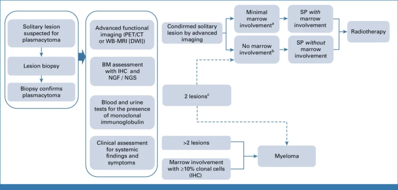
An SP requires histologic and immunohistochemical confirmation of a clonal plasma cell tumor, no additional lesions on advanced imaging, less than 10% clonal plasma cells in the bone marrow by immunohistochemistry, and no myeloma-defining events. Bone lesions with direct extension into adjacent soft tissue remain classified and managed as SBP, SEMP refers to a plasma cell tumor arising primarily at a nonosseous site.
The updated recommendations retain an important distinction between SP with and without minimal marrow involvement. Sensitive flow cytometry or sequencing may detect a small clonal population even when conventional marrow assessment shows no significant infiltration. These patients still meet the SP definition when clonal plasma cells remain below 10%, but the finding should be reported, because especially in SBP, it carries a greater risk of progression.
SPs can harbor many of the same cytogenetic and molecular abnormalities seen in MM, including hyperdiploidy, IgH rearrangements, del(13q), high-risk alterations such as del(17p), t(4;14), t(14;16), and 1q gain or amplification. In one SBP cohort, KRAS mutations and TP53 abnormalities were associated with inferior progression-free survival. These findings are prognostically interesting, but they don’t yet change the basic treatment approach.
Advanced Imaging Is Central to Diagnosis
A lesion cannot reliably be called solitary without whole-body imaging. Plain radiographs are insufficient, and conventional CT can miss early marrow disease. The IMWG recommends advanced functional imaging for every patient at diagnosis, preferably whole-body MRI with diffusion-weighted imaging or FDG-PET/CT. PET/CT is especially preferred for SEMP.
The effect of better imaging is clinically important. MRI identified focal or diffuse marrow involvement in more than 25% of patients previously classified as having SBP by conventional radiography. Small studies using PET/CT have found additional lesions in 33%-55% of patients initially considered to have SBP on plain radiographs. The most sensitive available modality should be used at baseline, and the same technique should generally be retained for subsequent response assessment.
Bone Marrow Assessment Has Become More Sensitive
Bone marrow aspirate alone without trephine immunohistochemistry or sensitive flow cytometry is considered inadequate. The IMWG recommends multiparametric or next-generation flow cytometry for both SBP and SEMP, with next-generation sequencing as an option when available. Cytogenetic evaluation by FISH or NGS in the marrow or plasmacytoma is also strongly recommended.
Sensitive assays can detect clonal marrow plasma cells in patients who would appear negative by conventional methods. In SBP, this finding has repeatedly been associated with earlier progression to MM: one recent cohort reported a median time to MM progression of 15.7 months in patients with marrow involvement compared with 79 months in those without it. Despite that, the IMWG does not currently recommend different initial treatment solely because minimal marrow disease is detected.
All patients should undergo evaluation for monoclonal immunoglobulin and free light chains before treatment and during follow-up. Small M-proteins are common in SBP and less frequent in SEMP. Serial measurements provide prognostic information when the monoclonal protein persists after local treatment.
Radiotherapy as the Mainstay of Treatment
Despite increasingly sensitive detection of minimal disease and the availability of effective MM drugs, definitive local radiotherapy remains the treatment of choice for SP. Reported response rates range from 83% to 96%. No randomized prospective study has established the optimal radiation dose.
The IMWG recommends 40-50 Gy in 20-25 fractions for SP. ILROG provides additional size-based guidance: 35-40 Gy for SBP smaller than 5 cm, with 35 Gy acceptable for small lesions, and 40-50 Gy for lesions 5 cm or larger. For SEMP, 40-50 Gy is recommended, with 40 Gy considered reasonable for small, well-defined tumors or after excision with positive margins. Palliative doses commonly used for plasmacytomas in established MM are inadequate when treating SP with curative intent.
IMRT and VMAT are preferred over conventional techniques, and ISRT should replace large historical fields. Routine regional lymph-node irradiation is not required for SEMP.
An unusual situation arises when imaging identifies two lesions without other evidence of systemic disease. The IMWG considers local treatment of both lesions with close surveillance, particularly when they are anatomically close and marrow evaluation is negative. Greater caution is advised when the lesions sit in separate regions or either exceeds 5 cm.
When Surgery Is Considered
Surgery has a limited but important role. In SBP, it may be required for vertebral instability, spinal cord compression, or an impending or established pathologic fracture. SEMP may occasionally be removed during diagnostic or therapeutic surgery.
Excision alone is generally inadequate because local recurrence is higher without radiotherapy. Postoperative RT should therefore be strongly considered, particularly when margins are positive or complete excision would require substantial functional sacrifice. Exceptionally, a small SEMP completely excised with adequate margins may be managed surgically.
Systemic Therapy Is Not Standard
A small nonrandomized study comparing IMRT alone with concurrent lenalidomide-dexamethasone reported 5-year myeloma-free survival of 100% with combined treatment versus 77.1% with RT alone, and PFS of 81.7% versus 48.4%.
Other retrospective studies have produced conflicting results and are difficult to interpret because systemic treatment was often preferentially given to patients with larger tumors, persistent M-protein, minimal marrow involvement, or inadequate imaging responses. Many older studies also lacked contemporary imaging and sensitive marrow assessment.
The IMWG therefore does not recommend adjuvant or concurrent systemic therapy when adequate radiotherapy can be delivered, regardless of tumor size or minimal marrow involvement.
Response Should Not Be Judged Too Early
Response assessment after SP radiotherapy requires patience. Radiation-related inflammation, marrow regeneration, bone remodeling, and residual fibrosis can persist after successful treatment and mimic active disease on imaging.
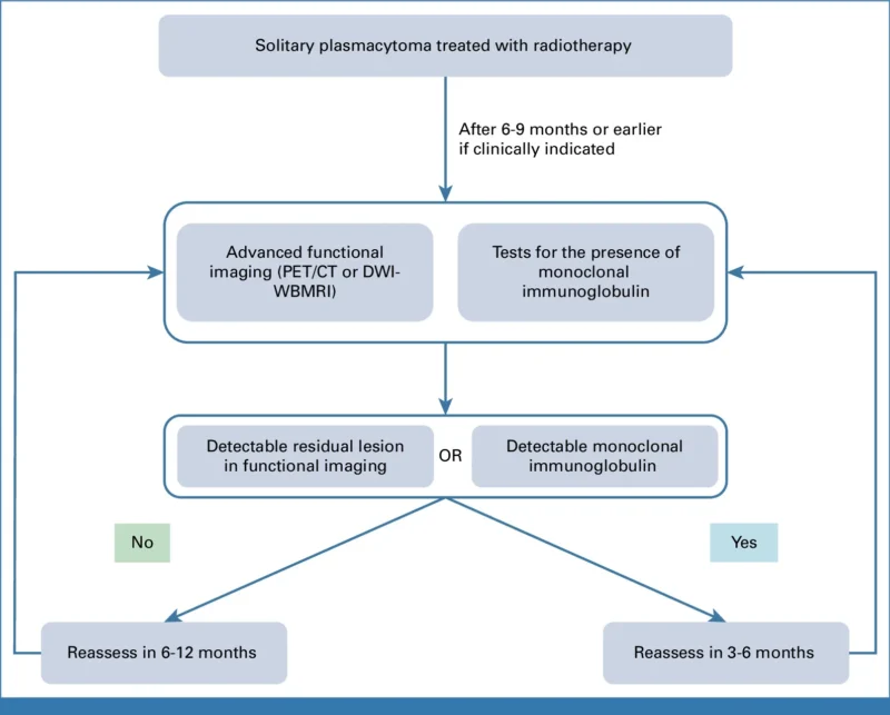
The IMWG recommends functional imaging with PET/CT or whole-body diffusion-weighted MRI 6-9 months after radiotherapy, using the same modality obtained at baseline and standardized IMPETUS or MY-RADS criteria. Imaging at around 3 months can be falsely positive.
For patients in imaging complete remission, annual functional imaging is suggested for the first 5 years. A residual or equivocal mass can be reassessed approximately every 6 months until complete response or stable residual abnormality is established.
Biochemical response follows a similar principle. M-protein may take months to disappear, and persistent M-protein is associated with increased risk of progression to MM. Monoclonal protein assessment should continue every 3-6 months, routine repeat bone marrow examinations are unnecessary unless clinically indicated.
Progression Risk Differs Between Bone and Extramedullary Disease
SBP carries the greater long-term risk of systemic progression. Approximately half of patients develop MM within 3-5 years after local radiotherapy, and reported 10-year progression rates reach 65%-84%. Even negative PET/CT or whole-body MRI at diagnosis does not eliminate this risk. Progression may appear as another focal lesion, multiple focal lesions with little marrow involvement, or conventional MM with diffuse marrow disease.
SEMP local recurrence occurs in approximately 10%, and reported progression to MM ranges from 11% to 36%. Recurrence can involve lymph nodes, skin or subcutaneous tissue, or new lytic bone lesions.
A new isolated lesion should prompt complete restaging before being considered systemic progression. A solitary recurrence outside the previous radiation field can still be treated with RT. For recurrence within or adjacent to a previously treated field, prior dose and treatment volumes must be reviewed before considering reirradiation. Apparent primary radioresistance is exceptionally uncommon.
Once a patient meets criteria for overt MM, standard systemic MM therapy applies. The IMWG specifically states that patients who progress from SP to MM should remain eligible for frontline MM clinical trials.
Solitary Plasmacytoma in 2026: Better Staging, Local Treatment, Longer Surveillance
The updated IMWG recommendations keep radiotherapy as the foundation of SP treatment, but substantially raise the bar for establishing that a disease is truly solitary in the first place. Whole-body functional imaging, sensitive marrow testing, and complete monoclonal protein assessment can now surface occult disease that older diagnostic approaches would have missed.
For patients who still meet SP criteria after that workup, management stays deliberately local: definitive modern RT, no routine adjuvant systemic therapy, and structured long-term surveillance. Minimal marrow disease and persistent M-protein sharpen the prognosis without yet giving clinicians grounds to convert a localized plasmacytoma into a systemic-treatment indication.
You can also read: