In their 2026 study published in Leukemia, Megan R. Daneman and colleagues identified experimental small molecules targeting acute myeloid leukemia (AML) by screening more than 4.2 million compounds using cheminformatics. These molecules kill AML cells by inhibiting glutathione reductase, raising oxidative stress and impairing mitochondrial function.
The compounds spared healthy cells in culture and acted synergistically with midostaurin and venetoclax. All of the work was done in cell lines and patient-derived cells, and none of the compounds has been tested in animals or patients.
The screen builds on the same group’s earlier work on the PS127 family of mitochondria-targeting compounds. It selects molecules by predicted function, and found active compounds whose structures differ substantially from the PS127 family.
Metabolic Vulnerabilities in AML
Relapse and drug resistance are major problems in AML. Induction chemotherapy reduces rapidly proliferating blasts, but leukemic stem cells may persist and cause recurrence. Both populations show metabolic features that could be targeted. AML cells often depend on oxidative phosphorylation and glutamine metabolism, and their high mitochondrial activity generates reactive oxygen species (ROS) that damage proteins, lipids and DNA. To limit this damage, they rely on antioxidant systems such as glutathione metabolism and on mechanisms that remove damaged mitochondria.
One such mechanism is mitophagy, the selective degradation of damaged mitochondria through autophagy. It normally supports survival, but excessive activation can contribute to cell death, and both stimulating and inhibiting mitophagy have been toxic to leukemia cells depending on context. The authors had previously found that PS127-family compounds disrupted mitochondrial bioenergetics and selectively killed AML cells, including leukemic stem cells, and suggested they promote apoptosis and interfere with redox-balancing enzymes. The new study aimed to find more compounds with these properties and to clarify how they work.
Screening 4.2 Million Compounds
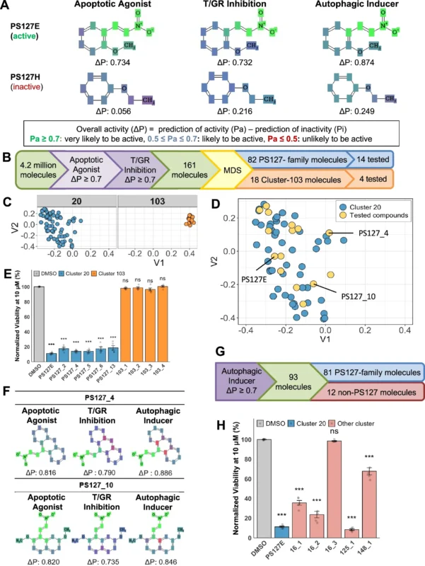
The investigators used Prediction of Activity Spectra for Substances (PASS), a platform that predicts biological activities from chemical structure. Instead of searching for binding to a predefined target, they selected compounds by predicted function. The first screen asked for two activities associated with PS127 compounds: promotion of apoptosis and inhibition of thioredoxin reductase or glutathione reductase. Stringent criteria gave 161 hits, which grouped into several structural clusters, one of them containing the original PS127 family.
Some compounds with both predicted activities did not kill AML cells. A third characteristic separated active from inactive molecules: the predicted ability to induce autophagy. Adding it cut the candidates from 161 to 93, of which 81 were related to the PS127 family and 12 were structurally distinct. Four of five structurally distinct candidates tested significantly reduced AML cell viability, so the combination of predicted activities can find active molecules outside a single chemical family.
Selective Activity Against AML Cells
Of 14 representative PS127-family compounds tested in AML cell lines, 12 had half-maximal cytotoxic concentrations below 8 μM, and four were submicromolar in MOLM-13 cells. Several were also active in MOLM-14, MV4;11, NB4 and OCI-AML2 cells, which represent different genetic backgrounds.
The compounds were generally more toxic to leukemic cells than to healthy cells. For the leading PS127 molecules, concentrations needed to kill healthy peripheral blood mononuclear cells were at least 12-fold higher than for MOLM-13 cells, and selected compounds were approximately 5- to 19-fold less toxic to cultured cardiomyocytes than to AML cells.
PS127E and PS127_10 also had substantially higher cytotoxic concentrations in healthy bone marrow-derived CD34-positive progenitors than in AML models. Mitochondrial dysfunction and oxidative stress also matter in normal tissues, and selectivity in cultured cells cannot establish the safety of prolonged treatment in vivo.
Glutathione Reductase as the Target
Flow cytometry confirmed that PS127E, PS127_4 and PS127_10 induced apoptosis. Combining them with autophagy inhibitors acting at different stages reduced cytotoxicity, so functional autophagy is needed for their full effect.
The screen had predicted inhibition of thioredoxin reductase or glutathione reductase, but the prioritized compounds did not significantly inhibit thioredoxin reductase. They inhibited glutathione reductase, the enzyme that converts oxidized glutathione back to its reduced, antioxidant form.
In AML cells, PS127-family compounds lowered the ratio of reduced to oxidized glutathione and significantly inhibited glutathione reductase activity, and this was not seen in healthy peripheral blood mononuclear cells under the same conditions. Genetic suppression of the glutathione reductase gene, GSR, also killed AML cells.
Supplementation with reduced glutathione or its precursor N-acetylcysteine rescued AML cell viability. Oxidized glutathione, ascorbate and α-tocopherol did not, so the toxicity appears tied to glutathione metabolism and is not readily offset by other antioxidants.
The compounds raised both cytosolic and mitochondrial ROS, and reduced glutathione reversed this. In thermal shift assays, active compounds bound purified glutathione reductase, with an estimated binding constant of 0.204 μM for PS127_10, consistent with high-affinity binding. The biochemical, genetic and rescue experiments together provide substantial evidence that glutathione reductase is a molecular target.
Mitochondrial Dysfunction and Lethal Mitophagy
After three hours, the prioritized compounds significantly reduced oxygen consumption in MOLM-13 cells, and intracellular ATP fell. Healthy peripheral blood mononuclear cells did not show the same reduction. The authors propose that glutathione reductase inhibition raises oxidative stress, damages mitochondria and activates mitophagy, which then contributes to cell death.
The rise in ROS, the impaired bioenergetics and the reduced cytotoxicity under autophagy inhibition support this, but the study does not establish the sequence from enzyme inhibition to lethal mitophagy, and the compounds may act on other targets. Transcriptomic and proteomic studies will be needed to define these effects.
A Structurally Different Compound
To test whether the screen works beyond the PS127 family, the researchers chose compound 125_1, which is structurally unrelated to the original leads but has the same three predicted activities. Its half-maximal cytotoxic concentration in MOLM-13 cells was approximately 0.41 μM, and it was more than 30-fold higher in healthy peripheral blood mononuclear cells and cultured cardiomyocytes.
It induced apoptosis, depended on autophagy, reduced glutathione reductase activity, raised cytosolic and mitochondrial ROS, bound glutathione reductase in thermal shift assays and impaired respiration and ATP production. Reduced glutathione rescued treated cells, and oxidized glutathione and other antioxidants did not.
Synergy With FLT3 Inhibitors and Venetoclax
The strongest interactions were with midostaurin, a multikinase inhibitor used in FLT3-mutated AML. PS127-family compounds were highly synergistic with it in FLT3-ITD-mutated MOLM-13 and MV4;11 cells. In MV4;11 cells, PS127_4 plus midostaurin reached an average maximum Bliss synergy score above 60. At one strongly synergistic combination, survival was approximately 13% in MV4;11 cells and 95% in healthy peripheral blood mononuclear cells.
Synergy also appeared in OCI-AML2 cells, which lack known FLT3 mutations, with varying magnitude, and the authors note that midostaurin has other targets that could contribute. The compounds were also synergistic with quizartinib, sorafenib and gilteritinib.
The authors propose that glutathione reductase inhibition acts as a metabolic sensitizer: by weakening the antioxidant defense of AML cells, it may increase their susceptibility to the mitochondrial disruption caused by FLT3 inhibition.
The precise mechanisms remain to be established. Strong synergy with venetoclax in MOLM-13 and MV4;11 cells may reflect the role of BCL-2 in mitochondrial survival and metabolic regulation, which could make glutathione reductase inhibition sensitize cells to venetoclax-induced apoptosis. Combinations with cytarabine and doxorubicin were generally less synergistic than those with midostaurin.
Patient-Derived AML Cells
Primary AML samples with and without FLT3 mutations were also tested. Mitochondrial oxygen consumption varied widely between samples. Even so, reduced glutathione rescued cells treated with PS127E or compound 125_1 in both FLT3-mutated and FLT3-wild-type samples.
PS127_10 combined with midostaurin or doxorubicin produced interactions from additive to strongly synergistic, depending on the sample, and FLT3 mutation status did not consistently predict the magnitude of synergy. The compounds may therefore exploit metabolic vulnerabilities across genotypes, but FLT3 status alone is unlikely to predict response.
Remaining Questions
The authors link the findings to leukemic stem cells. Earlier work showed that PS127B and PS127E induced apoptosis in leukemic stem cells and spared healthy hematopoietic stem cells. The CD34-positive progenitor data add to the evidence of selectivity, but they do not show the effect of the newly prioritized compounds on leukemic stem cells in patients.
The full range of compound interactions is uncharacterized, and RNA sequencing and proteomics could reveal further pathways behind efficacy or toxicity. A stable, highly resistant doxorubicin-resistant MOLM-13 population was not established, which limits conclusions about chemotherapy-resistant AML.
The authors also propose combinations with proteasome inhibitors and late-stage autophagy inhibitors, given the links between autophagy, protein homeostasis and leukemia cell survival. Animal studies, pharmacokinetics, off-target assessment and further medicinal chemistry are needed before the therapeutic potential can be judged.

Does 7+3 Still Hold Its Ground in AML? How Venetoclax Is Challenging Intensive Induction