

The Institute of Cancer Research shared a post on LinkedIn:
“We’ve shortlisted seven images for this year’s ICR’s Science and Medical Image competition, showcasing eye-catching science being carried out at the ICR.
Tell us your favourite in the comments.
ENTRY 1: A melanoma cell infected with an oncolytic herpesvirus (green), which causes syncytia — a single large cell containing multiple nuclei. A drug that disrupts cell division enhances this effect, making cancer cells more vulnerable to viral infection. This approach highlights a new strategy to boost oncolytic virotherapy by exploiting the dynamics of cancer cell division.
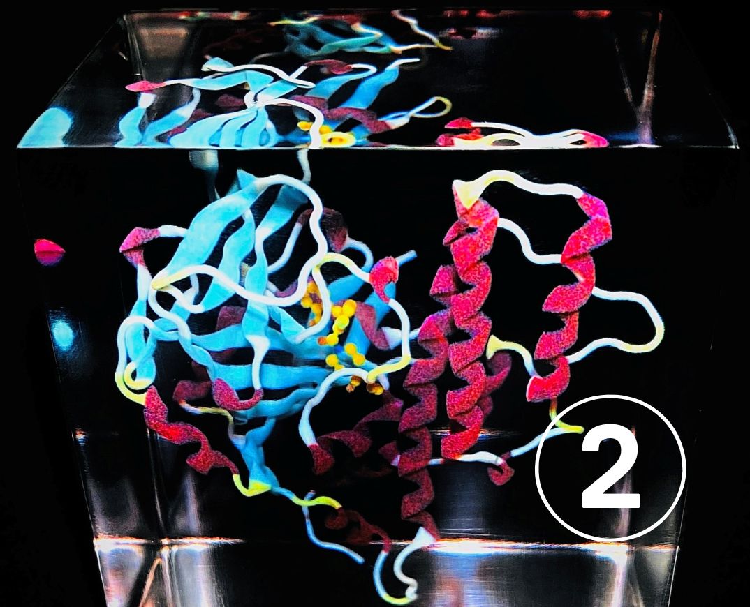
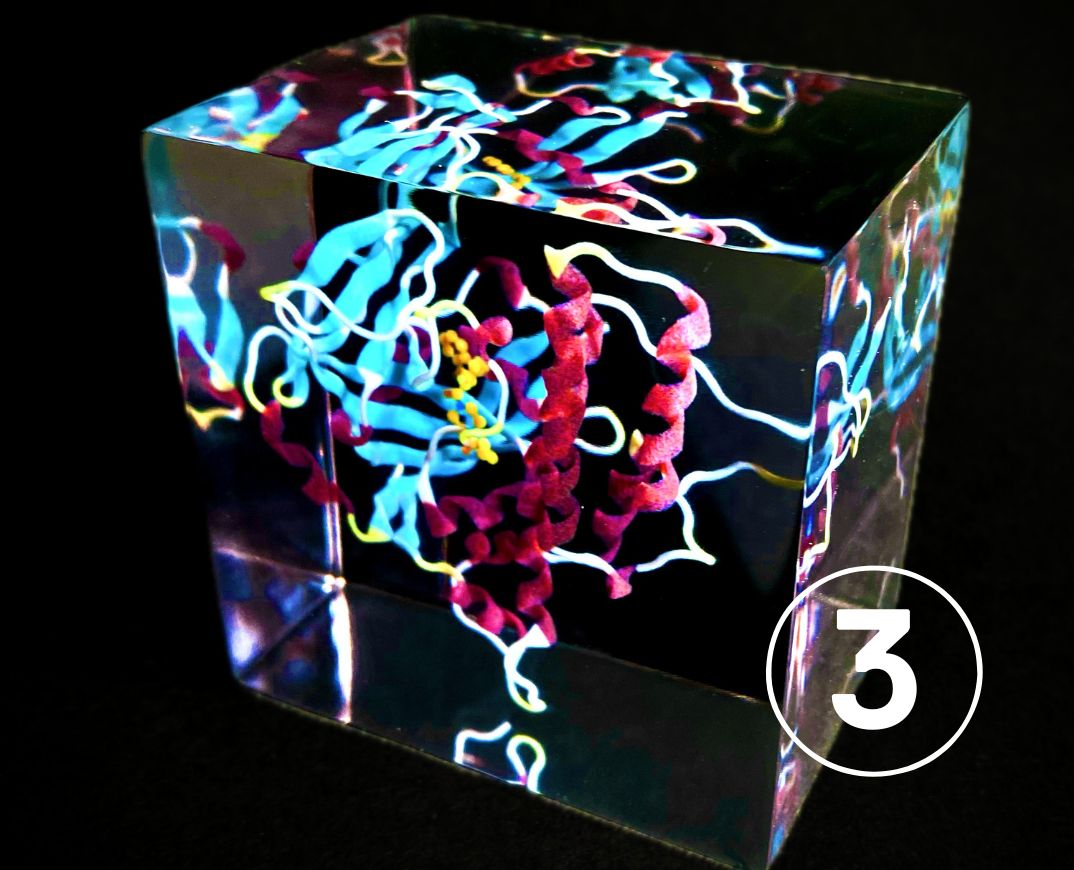
ENTRY 2 and 3: 3D printed model of the targeted drug Olaparib within the protein PARP1, used in the treatment of ovarian, breast, pancreatic, and prostate cancers. The drug binds at the ‘heart’ of the PARP1 protein and by coincidence the protein resembles a heart from various angles.
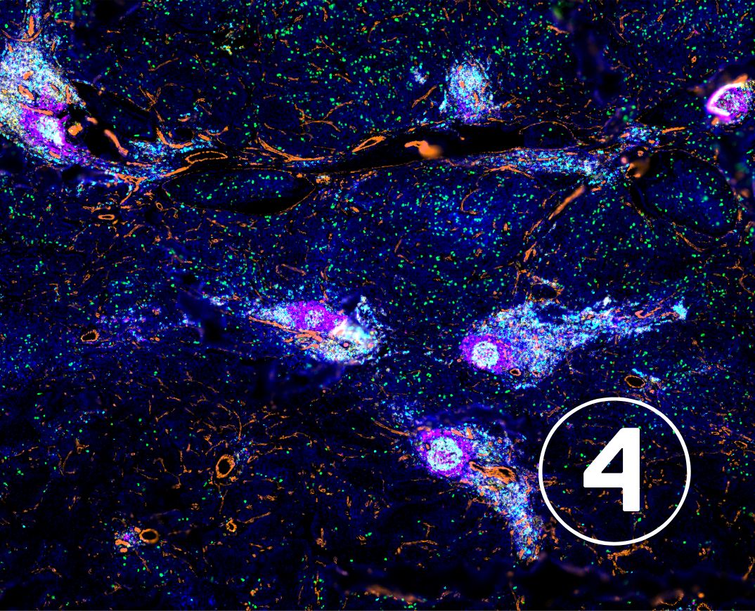
ENTRY 4: Immune cells forming aggregates in childhood Neuroblastoma tumour. B cells form round active antibody secreting structures surrounded by T cells (both helper and cytotoxic). The ability of uncover spatial relationship of immune cells within tumours provides crucial insights of their interaction and will gives us tools to identify new prognostic markers.
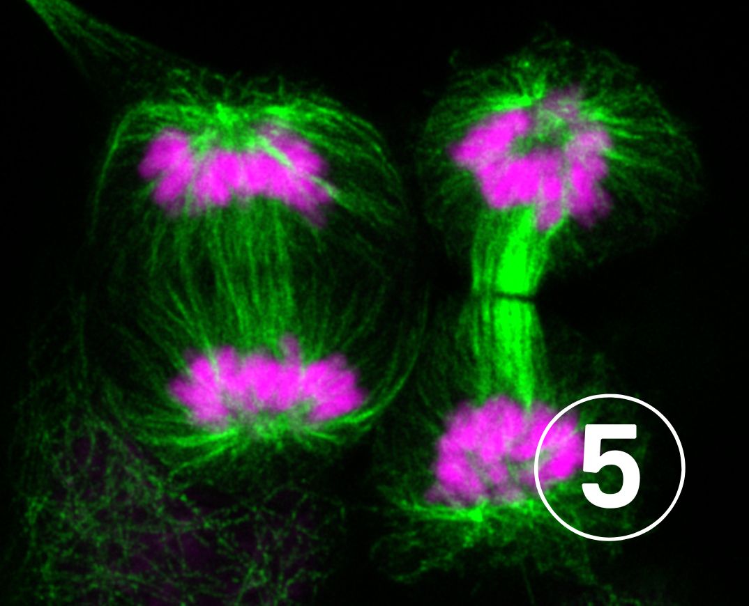
ENTRY 5: This image shows two cancer cells going through cell division – a process where one cell divides into two or more daughter cells. During cell division, narrow tube-like structures called microtubules (green) separate DNA (magenta) containing chromosomes and distribute them to daughter cells. Cells that divide in an uncontrolled manner and form tumors. Thus, stopping cancer cell division by drugs can be an effective strategy in the battle against cancer.
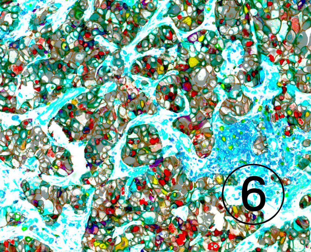
ENTRY 6: Using a technique known as Hyperplex Immunofluorescence, we can now visualise how various cell types are organised and interconnected within the tumour’s ecosystem. This technology offers valuable insights into how advanced prostate tumours evolve and adapt under treatment pressure.
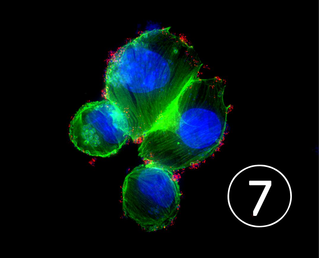
ENTRY 7: We engineered E. coli bacterium (shown in small blue dots) decorated with microscopic ‘stings’ (red) to deliver a cocktail of ‘poisons’ to pancreatic cancer cells, which completely takes over the pancreatic cancer cellular structure (green), forcing cancer cells to distort, shrink and die, and at the same time release signals to boost the immune system. We have found the efficiency of these engineered bacteria to eliminate pancreatic cancer cells and trigger beneficial immune response to be much superior than even high doses of available cancer therapy agents, and they could inspire the next generation of cancer therapy development.”

More posts featuring The Institute of Cancer Research.