
Retinoblastoma: What patients and caregivers should know about
What is Retinoblastoma?
Retinoblastoma is an eye cancer that typically develops in children before 5 years of age. This cancer develops in the retina—the part of the eye that helps a person see color and light. Retinoblastoma may affect one or both eyes. In about two-thirds of all cases, only one eye is affected 1.
Prevalence and Demographics
- Globally, retinoblastoma affects approximately 1 in 15,000 children, with an estimated 8,600 to 9,000 new cases each year2.
- The majority of these cases, about 90%, occur in developing countries, influenced by higher birth rates and larger proportions of young populations2.
- In developed countries like the U.S. and Europe, retinoblastoma accounts for around 3% of childhood cancers, while in some developing nations it can account for over 10%.
- The age-specific sex ratio tends to increase with age, with more males affected at older ages.
Types of Retinoblastoma: Heritable and Sporadic
Retinoblastoma can occur in two forms: heritable and sporadic.
1. Heritable Retinoblastoma:
-
- This form is caused by a germline mutation in the RB1 gene, which means the mutation can be passed from parent to child 3.
- Children with heritable retinoblastoma typically have mutations in all their cells, and they often develop tumors in both eyes (bilateral retinoblastoma)4.
- A small number of children with hereditary retinoblastoma develop cancers outside the eyes, such as in the pineal gland of the brain. Later in life, tumors might form in other parts of the body (bones, muscles, and skin).
Screening for Cancer in Children with Hereditary Retinoblastoma
-
- Eye Examinations: Starting possibly from birth, especially if there is a family history, children undergo regular eye exams conducted by an ophthalmologist. These exams are frequent (every one to two months) during the first two years and become less frequent as the child ages, transitioning to annual exams after five years1.
- MRI Scans: Due to the risk of developing tumors in the pineal gland, MRI scans of the brain are recommended twice a year until the age of four 1.
- Monitoring for Other Cancers: There is no standard screening for other cancers, but families are advised to be vigilant for unexplained symptoms like lumps, bumps, or persistent illness, which healthcare providers should evaluate1.
Prenatal testing
-
- Testing that occurs before pregnancy— Testing that happens before pregnancy is called preimplantation genetic testing (PGT). This special type of genetic testing is done along with in vitro fertilization (IVF). PGT offers a way to test embryos for a known RB1 mutation before placing them into the uterus5.
- Testing that occurs during pregnancy— Testing can be used to determine if a pregnancy is affected by a known RB1 mutation. A doctor gathers cells from the pregnancy in one of two ways 5
- Chorionic villus sampling (CVS) — During the first trimester
- Amniocentesis — During the second trimester or later
Once these tissues are collected, DNA is isolated and checked for the presence of the RB1 mutation identified in the family. Both of these tests carry minor risks and should be discussed with an experienced doctor and/or genetic counselor.
2. Sporadic Retinoblastoma:
-
- Most cases of retinoblastoma are sporadic, meaning the mutation in the RB1 gene occurs by chance after conception, affecting only the retinal cells6.
- Sporadic retinoblastoma, also known as non-hereditary retinoblastoma, primarily occurs due to mutations that happen in the retinal cells after conception, rather than being inherited from a parent. This form of retinoblastoma is typically unilateral, affecting only one eye, and does not have a familial pattern, meaning it generally does not run in families6.
- This type usually affects one eye (unilateral retinoblastoma) and is not passed down from the parents 6.
- Sporadic retinoblastoma is usually diagnosed at a later age compared to hereditary forms. The average age of diagnosis is around two years7.
- The mutations in sporadic retinoblastoma occur somatically, meaning they are acquired during the lifetime of the retinal cells and are not present in the germline. This type of mutation affects only the cells of the retina where the mutation occurs and is not found in all cells of the body.
- Children with sporadic retinoblastoma do not typically pass the condition to their offspring because the mutations are not present in their germline cells1.
- Unlike hereditary retinoblastoma, children with sporadic retinoblastoma have a significantly lower risk of developing secondary cancers later in life.
- The diagnosis of sporadic retinoblastoma involves a detailed eye examination, imaging tests such as ultrasound, MRI, or CT scans, and possibly genetic testing to confirm the absence of germline RB1 mutations8.
- Unlike hereditary retinoblastoma, where genetic testing can identify at-risk individuals, sporadic retinoblastoma does not have a known genetic marker for screening. Therefore, genetic testing is not a tool for screening in sporadic cases.
Symptoms
Common Symptoms
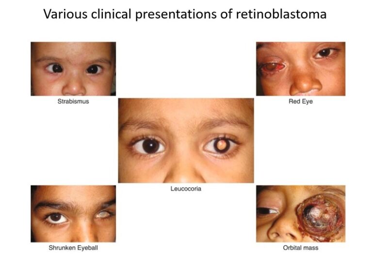
- Leukocoria (White Pupil):
- The most common and noticeable symptom of retinoblastoma is leukocoria, where the pupil appears white or has a white reflection when light shines on it, especially noticeable in photographs taken with a flash9.
- Strabismus (Crossed Eyes):
- Misalignment of the eyes, where one eye may not look in the same direction as the other, is often referred to as a “crossed eye” or “lazy eye”10.
- Vision Problems:
- Affected children may have poor vision, which might be noticed if the child has difficulty seeing or is unusually clumsy9.
- Eye Redness and Swelling:
- Redness in the white part of the eye and swelling around the eye are also common symptoms9.
Less Common Symptoms
- Eye Pain:
- Some children may experience pain in the affected eye11.
- Different-Colored Irises:
- A change in the color of the iris or different-colored irises may occur10.
- Enlarged Pupil:
- The pupil may appear larger or does not constrict in response to light10.
Advanced Symptoms
- Deterioration in Appearance of the Eye:
- The eye may appear larger, or there might be cloudiness in the front part of the eye11.
- Systemic Symptoms:
- If the cancer spreads beyond the eye, symptoms can include loss of appetite, weight loss, headaches, vomiting, and bone pain if metastasis occurs to the bones11.
Causes
While the exact cause of the RB1 mutation in sporadic cases is not well understood, certain genetic and environmental factors may contribute to the risk. Children with a family history of retinoblastoma are at a higher risk, especially if they inherit a faulty copy of the RB1 gene from one of their parents. However, most cases appear without any previous family history of the disease.
The primary cause of retinoblastoma is mutations in the RB1 gene, a tumor suppressor gene. This gene plays a critical role in regulating cell growth by preventing cells from dividing too quickly or uncontrollably. When the RB1 gene functions correctly, it produces a protein that helps control the cell cycle, acting as a brake on cell division. However, if a mutation occurs in the RB1 gene, this control is lost, leading to rapid and uncontrolled cell growth, resulting in cancer3.
Diagnosis
The diagnosis of retinoblastoma involves a combination of clinical examination and various imaging and diagnostic tests. Here is a brief overview of the diagnostic approaches12,13,14:
Clinical Examination
- Physical Examination:
- A thorough eye examination is crucial. This includes checking for signs like leukocoria (white pupil), strabismus (misaligned eyes), and other visible abnormalities in the eye.
- An examination under anesthesia is often necessary for a detailed assessment, especially in young children who may not cooperate with an eye exam.
- Fundus Examination:
- A dilated fundus examination allows the ophthalmologist to view the retina directly and look for any tumors or abnormalities. Retinoblastoma typically presents as a white or cream-colored mass in the retina, often with increased vascularization.
Imaging Tests
- Ultrasound:
- Ultrasound is a primary imaging tool used to confirm the presence of a mass within the eye and can provide details about the size and extent of the tumor.
- Magnetic Resonance Imaging (MRI):
- MRI is highly valuable for assessing the extent of the tumor, particularly whether it has spread beyond the eye. It provides detailed images of the soft tissues around the eye and can help in staging the cancer.
")
Axial T2-weighted MRI of the brain and orbit showing retinoblastoma in the left eye along with retinal detachment (red arrow)
- MRI is highly valuable for assessing the extent of the tumor, particularly whether it has spread beyond the eye. It provides detailed images of the soft tissues around the eye and can help in staging the cancer.
- Computed Tomography (CT) Scan:
- CT scans are less commonly used due to the risk of radiation but can help detect calcium deposits within the tumor, which is characteristic of retinoblastoma.
- Fundus Photography:
- This involves taking detailed photographs of the retina to document the appearance of the tumor and is useful for monitoring the tumor over time.
Additional Diagnostic Tests
- Fluorescein Angiography:
- This test involves injecting a dye into the bloodstream and taking photographs of the retina as the dye passes through the blood vessels. It helps in identifying any abnormal blood vessels associated with the tumor.
- Optical Coherence Tomography (OCT):
- OCT provides high-resolution images of the retina and can be used to observe the tumor’s effect on the retinal structure.
Genetic Testing
- Genetic Testing:
- As stated above for children diagnosed with retinoblastoma, genetic testing is recommended to determine if the tumor is heritable, which involves mutations in the RB1 gene.
Biopsy
Typically, a biopsy is not performed in cases of retinoblastoma due to the risk of spreading the tumor cells15. The diagnosis is usually made based on clinical examination and imaging findings.
Importance of Early Detection
Early detection of these symptoms is crucial for effective treatment16. Parents and caregivers are often the first to notice signs like leukocoria in photographs. Healthcare providers emphasize the importance of regular pediatric check-ups that can identify early signs of retinoblastoma, potentially before it is visible to the untrained eye17. If any of these symptoms are observed, it is important to consult a healthcare provider promptly to determine the cause and, if necessary, begin appropriate treatment. Early diagnosis and treatment are key to improving outcomes for children with retinoblastoma18.
Treatment
Standard Treatment Modalities
- Chemotherapy:
- Systemic Chemotherapy: Traditionally involves drugs like vincristine, etoposide, and carboplatin. These are used to shrink tumors, making them amenable to focal therapies19.
- Intra-arterial Chemotherapy (IAC): Delivers chemotherapy directly to the ophthalmic artery, minimizing systemic toxicity and enhancing drug delivery to the tumor20. The 1-year Kaplan–Meier estimate for ocular survival after IAC was 96% across different groups. Another study comparing intravenous chemotherapy (IVC) and IAC found the overall success rate was higher with IAC than with IVC (75.7% vs. 69.5%)21. There has been a long controversy over intra-arterial chemo for retinoblastoma – Guillermo Chantada
- Intravitreal Chemotherapy: Used for tumors with vitreous seeding, involves direct injection into the vitreous cavity to control local tumor spread.
- Local Therapies:
- Cryotherapy: Utilizes extreme cold to destroy tumor cells, effective for smaller peripheral tumors. .
- Laser Therapy (Thermotherapy): Uses heat to destroy cancer cells. It’s often used in conjunction with chemotherapy to treat smaller tumors11. Effective for tumors ≤ 3 disc diameters (DD) regardless of prior chemotherapy.
- Brachytherapy: Involves placing radioactive plaques close to the tumor, suitable for medium-sized tumors located away from critical structures22.
- Enucleation: Removal of the eye is considered when the tumor is too large to treat with eye-conserving methods or if there is little hope of retaining useful vision. This method ensures the complete removal of the tumor23.
Recent Advances and Experimental Approaches
- Targeted Therapy and Immunotherapy:
- Research is ongoing into targeted therapies that exploit genetic changes in retinoblastoma cells. For example, VCN-01, an oncolytic virus, targets cells lacking the functional RB1 gene, showing promise in early clinical trials24.
- Immunotherapeutic approaches like GD2-targeted antibodies are under investigation, aiming to harness the body’s immune system to fight the tumor.
- Novel Drug Delivery Systems:
- Suprachoroidal Drug Delivery: This method involves injecting chemotherapy drugs into the suprachoroidal space, potentially reducing systemic toxicity and improving drug concentration at the tumor site25.
- Nanoparticle-mediated Therapy: Explores the use of nanoparticles to deliver chemotherapy directly to tumor cells, enhancing efficacy and reducing side effects26.
Multidisciplinary Approach
Treatment planning typically involves a multidisciplinary team including pediatric oncologists, ophthalmologists, radiation oncologists, and geneticists. This team tailors the treatment plan based on the tumor’s size, location, genetic makeup, and the patient’s overall health.
Supportive Care and Monitoring
Long-term monitoring is crucial for detecting recurrences and managing late effects of treatment, such as second malignancies and ocular complications. Patients often require lifelong surveillance.
Prognosis
The prognosis for retinoblastoma is generally favorable, with a survival rate of nearly 100% in the United States when appropriately treated. However, the prognosis can vary based on the cancer’s stage at diagnosis and the presence of metastasis. Early detection and treatment are crucial for preserving vision and improving survival outcomes27,28. Doctors use the stage of retinoblastoma to choose treatment and predict the outlook for a child. Three staging systems might be used for retinoblastoma29
The International Classification for Intraocular Retinoblastoma uses groupings to help predict the likelihood that the eye can be saved with current treatment options. Children in group A have the most favorable prognosis and are most likely to be able to see after treatment.
The Reese-Ellsworth staging system also uses groupings to help predict whether the child will be able to see after treatment. Children in group 1 (the very favorable stage) are most likely to be able to see after treatment.
The TNM staging system is based on tumor size and spread. The lower the stage of retinoblastoma, the better the expected outcome.
Watch the survival story of Rubi
Patient’s Survivorship
Survivors of retinoblastoma, particularly those with the hereditary form of the disease, face several long-term complications and health risks after their initial treatment. These complications can significantly impact their quality of life and require ongoing medical surveillance and care. Here are the key complications and risks based on the provided sources30,31,32:
Physical Health Complications
- Second Primary Cancers: Survivors, especially those treated with radiation, have a heightened risk of developing second primary cancers. Common types include osteosarcoma (bone cancer), soft tissue sarcomas, and melanoma of the skin.
- Vision Problems: Many survivors experience vision issues, ranging from partial to total blindness in the affected eye(s), depending on the extent and location of the tumors and the type of treatment received.
- Other Health Issues: Depending on the treatment modalities used (such as radiation and chemotherapy), survivors may also face a range of other health problems including cataracts, reduced kidney function, heart problems, and issues related to bone growth around the eye.
Psychological and Social Complications
- Mental Health Challenges: Many survivors deal with psychological impacts such as anxiety, depression, and post-traumatic stress disorder (PTSD), which can stem from their early cancer experiences and ongoing health challenges.
- Social and Functional Challenges: Vision issues can affect survivors’ social interactions, educational progress, and overall ability to perform daily activities. This can lead to challenges in social integration and educational attainment.
Challenges in Accessing Care
- Knowledge Gaps Among Healthcare Providers: There is often a lack of familiarity with the long-term risks associated with retinoblastoma among general healthcare providers, which can complicate survivors’ efforts to receive appropriate follow-up care.
- Need for Self-Advocacy: Survivors frequently find themselves needing to advocate for appropriate care and educate their healthcare providers about their unique health risks and needs.
What Patients Should Do
Long-Term Follow-Up Care
- Children treated for retinoblastoma should have life-long, follow-up care to monitor for any recurrence or late effects of treatment31
- Follow-up visits will focus on enhancing the child’s quality of life, addressing developmental and emotional concerns, and checking for signs of secondary tumors, especially in those with the genetic form of retinoblastoma.
Eye Care and Rehabilitation
- Patients treated with external beam radiation therapy may experience eye complications like cataracts, dry eyes, or vision problems, requiring regular ophthalmologic monitoring and management.
- Those who had an eye removed (enucleation) need a well-fitted prosthetic eye that is changed over time as the child grows. Special education services may be required for children who lost both eyes31
Monitoring for Recurrence and New Cancers
- Periodic imaging tests (CT/MRI scans) and blood tests may be done to check for any recurrence of retinoblastoma.
- Patients with the genetic form and those treated with radiation have an increased risk of developing other cancers later in life, so they require careful monitoring and screening.
General Precautions
- Wear sunglasses and protective eyewear to prevent further eye damage32
- Avoid activities that may injure the eyes, like playing with sharp toys or fireworks.
- Maintain regular follow-up visits with ophthalmologists and oncologists experienced in retinoblastoma care.
Watch the video, in which Carol L. Shields, MD, Co-Director of the Wills Eye Oncology Service in Philadelphia discusses Retinoblastoma
Conclusion
While retinoblastoma poses significant challenges, the prognosis is generally favorable with early detection and treatment. Lifelong surveillance and support are necessary to address the ongoing needs of survivors and enhance their quality of life.
References and useful resources
Clinicaltrials.gov – a huge database of clinical trials, useful for patients and families to find past, ongoing, and upcoming clinical trials.
Cancer.Net – Comprehensive information for people with cancer, families, and caregivers, from the American Society of Clinical Oncology (ASCO)
Cancer.Gov – More comprehensive overview on the treatment of retinoblastoma by NCI
Chop.edu – Children’s Hospital of Philadelphia
Cancer.org – American Cancer Society
Oncodaily.com – Online platform where you can find anything related to cancer such as everyday news, blogs, videos, podcasts, etc.